+Open data
-Basic information
Entry | Database: PDB / ID: 3j82 | ||||||
---|---|---|---|---|---|---|---|
Title | Electron cryo-microscopy of DNGR-1 in complex with F-actin | ||||||
Components |
| ||||||
Keywords | MEMBRANE PROTEIN/ADP-BINDING PROTEIN / DNGR-1 / Actin / Recognition of damage-associated molecular patterns / MEMBRANE PROTEIN-ADP-BINDING PROTEIN complex | ||||||
Function / homology | Function and homology information positive regulation of cytokine production => GO:0001819 / positive regulation of norepinephrine uptake / cellular response to cytochalasin B / regulation of transepithelial transport / morphogenesis of a polarized epithelium / bBAF complex / postsynaptic actin cytoskeleton organization / npBAF complex / nBAF complex / protein localization to adherens junction ...positive regulation of cytokine production => GO:0001819 / positive regulation of norepinephrine uptake / cellular response to cytochalasin B / regulation of transepithelial transport / morphogenesis of a polarized epithelium / bBAF complex / postsynaptic actin cytoskeleton organization / npBAF complex / nBAF complex / protein localization to adherens junction / brahma complex / postsynaptic actin cytoskeleton / Tat protein binding / structural constituent of postsynaptic actin cytoskeleton / GBAF complex / dense body / Formation of annular gap junctions / regulation of G0 to G1 transition / Gap junction degradation / Cell-extracellular matrix interactions / Folding of actin by CCT/TriC / apical protein localization / regulation of double-strand break repair / adherens junction assembly / regulation of nucleotide-excision repair / Prefoldin mediated transfer of substrate to CCT/TriC / RSC-type complex / RHOF GTPase cycle / Regulation of MITF-M-dependent genes involved in pigmentation / Adherens junctions interactions / tight junction / Sensory processing of sound by outer hair cells of the cochlea / regulation of norepinephrine uptake / Interaction between L1 and Ankyrins / Sensory processing of sound by inner hair cells of the cochlea / regulation of mitotic metaphase/anaphase transition / SWI/SNF complex / positive regulation of double-strand break repair / regulation of synaptic vesicle endocytosis / positive regulation of T cell differentiation / apical junction complex / establishment or maintenance of cell polarity / regulation of cyclin-dependent protein serine/threonine kinase activity / maintenance of blood-brain barrier / cortical cytoskeleton / positive regulation of stem cell population maintenance / NuA4 histone acetyltransferase complex / nitric-oxide synthase binding / regulation of G1/S transition of mitotic cell cycle / Recycling pathway of L1 / kinesin binding / brush border / calyx of Held / negative regulation of cell differentiation / positive regulation of double-strand break repair via homologous recombination / EPH-ephrin mediated repulsion of cells / RHO GTPases Activate WASPs and WAVEs / RHO GTPases activate IQGAPs / regulation of protein localization to plasma membrane / positive regulation of myoblast differentiation / EPHB-mediated forward signaling / substantia nigra development / receptor-mediated endocytosis / axonogenesis / negative regulation of protein binding / Translocation of SLC2A4 (GLUT4) to the plasma membrane / actin filament / cell motility / RHO GTPases Activate Formins / positive regulation of cell differentiation / adherens junction / FCGR3A-mediated phagocytosis / regulation of transmembrane transporter activity / Hydrolases; Acting on acid anhydrides; Acting on acid anhydrides to facilitate cellular and subcellular movement / Signaling by high-kinase activity BRAF mutants / DNA Damage Recognition in GG-NER / MAP2K and MAPK activation / B-WICH complex positively regulates rRNA expression / tau protein binding / Schaffer collateral - CA1 synapse / structural constituent of cytoskeleton / kinetochore / Regulation of actin dynamics for phagocytic cup formation / platelet aggregation / VEGFA-VEGFR2 Pathway / nuclear matrix / cytoplasmic ribonucleoprotein granule / Signaling by RAF1 mutants / Signaling by moderate kinase activity BRAF mutants / Paradoxical activation of RAF signaling by kinase inactive BRAF / Signaling downstream of RAS mutants / UCH proteinases / nucleosome / Signaling by BRAF and RAF1 fusions / cell-cell junction / Clathrin-mediated endocytosis / actin cytoskeleton / Factors involved in megakaryocyte development and platelet production / lamellipodium / presynapse Similarity search - Function | ||||||
Biological species | Mus musculus (house mouse) Homo sapiens (human) | ||||||
Method | ELECTRON MICROSCOPY / single particle reconstruction / cryo EM / Resolution: 7.7 Å | ||||||
Authors | Hanc, P. / Fujii, T. / Yamada, Y. / Huotari, J. / Schulz, O. / Ahrens, S. / Kjaer, S. / Way, M. / Namba, K. / Reis e Sousa, C. | ||||||
Citation | 
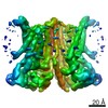
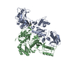
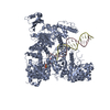
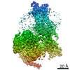
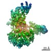
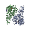
Journal: Immunity / Year: 2015 Title: Structure of the Complex of F-Actin and DNGR-1, a C-Type Lectin Receptor Involved in Dendritic Cell Cross-Presentation of Dead Cell-Associated Antigens. Authors: Pavel Hanč / Takashi Fujii / Salvador Iborra / Yurika Yamada / Jatta Huotari / Oliver Schulz / Susan Ahrens / Svend Kjær / Michael Way / David Sancho / Keiichi Namba / Caetano Reis e Sousa / Abstract: DNGR-1 is a C-type lectin receptor that binds F-actin exposed by dying cells and facilitates cross-presentation of dead cell-associated antigens by dendritic cells. Here we present the structure of ...DNGR-1 is a C-type lectin receptor that binds F-actin exposed by dying cells and facilitates cross-presentation of dead cell-associated antigens by dendritic cells. Here we present the structure of DNGR-1 bound to F-actin at 7.7 Å resolution. Unusually for F-actin binding proteins, the DNGR-1 ligand binding domain contacts three actin subunits helically arranged in the actin filament, bridging over two protofilaments, as well as two neighboring actin subunits along one protofilament. Mutation of residues predicted to mediate ligand binding led to loss of DNGR-1-dependent cross-presentation of dead cell-associated antigens, formally demonstrating that the latter depends on F-actin recognition. Notably, DNGR-1 has relatively modest affinity for F-actin but multivalent interactions allow a marked increase in binding strength. Our findings shed light on modes of actin binding by cellular proteins and reveal how extracellular detection of cytoskeletal components by dedicated receptors allows immune monitoring of loss of cellular integrity. | ||||||
History |
|
-Structure visualization
Movie |
Movie viewer |
---|---|
Structure viewer | Molecule: MolmilJmol/JSmol |
-Downloads & links
-Download
PDBx/mmCIF format | 3j82.cif.gz | 215.8 KB | Display | PDBx/mmCIF format |
---|---|---|---|---|
PDB format | pdb3j82.ent.gz | 175.9 KB | Display | PDB format |
PDBx/mmJSON format | 3j82.json.gz | Tree view | PDBx/mmJSON format | |
Others | Other downloads |
-Validation report
Summary document | 3j82_validation.pdf.gz | 959.9 KB | Display | wwPDB validaton report |
---|---|---|---|---|
Full document | 3j82_full_validation.pdf.gz | 1.1 MB | Display | |
Data in XML | 3j82_validation.xml.gz | 67.3 KB | Display | |
Data in CIF | 3j82_validation.cif.gz | 95.1 KB | Display | |
Arichive directory | https://data.pdbj.org/pub/pdb/validation_reports/j8/3j82 ftp://data.pdbj.org/pub/pdb/validation_reports/j8/3j82 | HTTPS FTP |
-Related structure data
Related structure data | 6102MC M: map data used to model this data C: citing same article (ref.) |
---|---|
Similar structure data |
-Links
-Assembly
Deposited unit |
|
---|---|
1 |
|
-Components
#1: Protein | Mass: 14968.989 Da / Num. of mol.: 1 Source method: isolated from a genetically manipulated source Source: (gene. exp.) Mus musculus (house mouse) / Gene: Clec9a, Dngr-1 / Production host: Homo sapiens (human) / References: UniProt: Q8BRU4 | ||||
---|---|---|---|---|---|
#2: Protein | Mass: 41664.484 Da / Num. of mol.: 3 / Source method: isolated from a natural source / Details: Human platelet actin / Source: (natural) Homo sapiens (human) / References: UniProt: P60709 #3: Chemical | ChemComp-CA / | #4: Chemical | |
-Experimental details
-Experiment
Experiment | Method: ELECTRON MICROSCOPY |
---|---|
EM experiment | Aggregation state: FILAMENT / 3D reconstruction method: single particle reconstruction |
-Sample preparation
Component |
| ||||||||||||||||
---|---|---|---|---|---|---|---|---|---|---|---|---|---|---|---|---|---|
Buffer solution | pH: 7.5 | ||||||||||||||||
Specimen | Embedding applied: NO / Shadowing applied: NO / Staining applied: NO / Vitrification applied: YES | ||||||||||||||||
Specimen support | Details: R0.6/1.0, Quantifoil | ||||||||||||||||
Vitrification | Instrument: FEI VITROBOT MARK II / Cryogen name: ETHANE / Humidity: 90 % |
-Electron microscopy imaging
Microscopy | Model: JEOL 3200FSC / Date: Dec 10, 2012 |
---|---|
Electron gun | Electron source: FIELD EMISSION GUN / Accelerating voltage: 200 kV / Illumination mode: FLOOD BEAM |
Electron lens | Mode: BRIGHT FIELD / Nominal magnification: 60000 X / Calibrated magnification: 109489 X / Nominal defocus max: 2000 nm / Nominal defocus min: 1000 nm / Cs: 1.6 mm / Camera length: 0 mm |
Specimen holder | Specimen holder model: JEOL 3200FSC CRYOHOLDER / Temperature: 55 K / Temperature (max): 60 K / Temperature (min): 50 K / Tilt angle max: 0 ° / Tilt angle min: 0 ° |
Image recording | Electron dose: 20 e/Å2 / Film or detector model: TVIPS TEMCAM-F416 (4k x 4k) |
EM imaging optics | Energyfilter name: JEOL Omega filter / Energyfilter upper: 20 eV / Energyfilter lower: 0 eV |
-Processing
EM software |
| ||||||||||||
---|---|---|---|---|---|---|---|---|---|---|---|---|---|
CTF correction | Details: Each Particle | ||||||||||||
Helical symmerty | Angular rotation/subunit: 166.6 ° / Axial rise/subunit: 27.6 Å / Axial symmetry: C1 | ||||||||||||
3D reconstruction | Method: IHRSR / Resolution: 7.7 Å / Resolution method: FSC 0.143 CUT-OFF / Num. of particles: 73608 / Details: (Single particle--Applied symmetry: C1) / Symmetry type: HELICAL | ||||||||||||
Refinement step | Cycle: LAST
|