 Movie
Movie Controller

[English] 日本語
 Yorodumi
- PDB-3j7w: Capsid Expansion Mechanism Of Bacteriophage T7 Revealed By Multi-... -
+ Open data

- Basic information
| Entry | Database: PDB / ID: 3j7w | ||||||
|---|---|---|---|---|---|---|---|
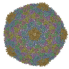
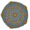
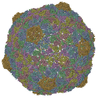
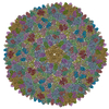
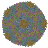
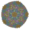
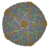
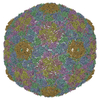
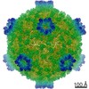
| Title | Capsid Expansion Mechanism Of Bacteriophage T7 Revealed By Multi-State Atomic Models Derived From Cryo-EM Reconstructions | ||||||
 Components Components | Major capsid protein 10A | ||||||
 Keywords Keywords |  VIRUS / maturation / DNA packaging / Procapsid / Non-covalent topological linking VIRUS / maturation / DNA packaging / Procapsid / Non-covalent topological linking | ||||||
| Function / homology | Capsid Gp10A/Gp10B / : / Major capsid protein / viral capsid / identical protein binding / Major capsid protein Function and homology information Function and homology information | ||||||
| Biological species |   Enterobacteria phage T7 (virus) Enterobacteria phage T7 (virus) | ||||||
| Method | ELECTRON MICROSCOPY / single particle reconstruction / cryo EM / Resolution: 3.5 Å | ||||||
 Authors Authors | Guo, F. / Liu, Z. / Fang, P.A. / Zhang, Q. / Wright, E.T. / Wu, W. / Zhang, C. / Vago, F. / Ren, Y. / Jakata, J. ...Guo, F. / Liu, Z. / Fang, P.A. / Zhang, Q. / Wright, E.T. / Wu, W. / Zhang, C. / Vago, F. / Ren, Y. / Jakata, J. / Chiu, W. / Serwer, P. / Jiang, W. | ||||||
 Citation Citation | Journal: Proc Natl Acad Sci U S A / Year: 2014 Title: Capsid expansion mechanism of bacteriophage T7 revealed by multistate atomic models derived from cryo-EM reconstructions. Authors: Fei Guo / Zheng Liu / Ping-An Fang / Qinfen Zhang / Elena T Wright / Weimin Wu / Ci Zhang / Frank Vago / Yue Ren / Joanita Jakana / Wah Chiu / Philip Serwer / Wen Jiang /  Abstract: Many dsDNA viruses first assemble a DNA-free procapsid, using a scaffolding protein-dependent process. The procapsid, then, undergoes dramatic conformational maturation while packaging DNA. For ...Many dsDNA viruses first assemble a DNA-free procapsid, using a scaffolding protein-dependent process. The procapsid, then, undergoes dramatic conformational maturation while packaging DNA. For bacteriophage T7 we report the following four single-particle cryo-EM 3D reconstructions and the derived atomic models: procapsid (4.6-Å resolution), an early-stage DNA packaging intermediate (3.5 Å), a later-stage packaging intermediate (6.6 Å), and the final infectious phage (3.6 Å). In the procapsid, the N terminus of the major capsid protein, gp10, has a six-turn helix at the inner surface of the shell, where each skewed hexamer of gp10 interacts with two scaffolding proteins. With the exit of scaffolding proteins during maturation the gp10 N-terminal helix unfolds and swings through the capsid shell to the outer surface. The refolded N-terminal region has a hairpin that forms a novel noncovalent, joint-like, intercapsomeric interaction with a pocket formed during shell expansion. These large conformational changes also result in a new noncovalent, intracapsomeric topological linking. Both interactions further stabilize the capsids by interlocking all pentameric and hexameric capsomeres in both DNA packaging intermediate and phage. Although the final phage shell has nearly identical structure to the shell of the DNA-free intermediate, surprisingly we found that the icosahedral faces of the phage are slightly (∼4 Å) contracted relative to the faces of the intermediate, despite the internal pressure from the densely packaged DNA genome. These structures provide a basis for understanding the capsid maturation process during DNA packaging that is essential for large numbers of dsDNA viruses. | ||||||
| History |
|
- Structure visualization
| Movie |
Movie viewer |
|---|---|
| Structure viewer | Molecule: MolmilJmol/JSmol |
- Downloads & links
-Download
| PDBx/mmCIF format | 3j7w.cif.gz | 444.2 KB | Display | PDBx/mmCIF format |
|---|---|---|---|---|
| PDB format | pdb3j7w.ent.gz | 366.5 KB | Display | PDB format |
| PDBx/mmJSON format | 3j7w.json.gz | Tree view | PDBx/mmJSON format | |
| Others |  Other downloads Other downloads |
-Validation report
| Arichive directory | https://data.pdbj.org/pub/pdb/validation_reports/j7/3j7wftp://data.pdbj.org/pub/pdb/validation_reports/j7/3j7w | HTTPS FTP |
|---|
-Related structure data
| Related structure data | 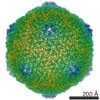 6035MC 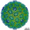 6034C 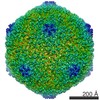 6036C 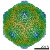 6037C 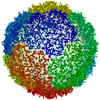 3j7vC 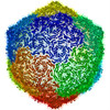 3j7xC C: citing same article ( M: map data used to model this data |
|---|---|
| Similar structure data |

-Links
 PDBj
PDBj- Assembly
| Deposited unit | 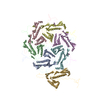
|
|---|---|
| 1 | x 60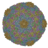
|
| 2 |
|
| 3 | x 5
|
| 4 | x 6
|
| 5 | 
|
| Symmetry | Point symmetry: (Schoenflies symbol: I (icosahedral)) |
-Components
| #1: Protein | Mass: 36589.625 Da / Num. of mol.: 7 / Source method: isolated from a natural source / Source: (natural) Enterobacteria phage T7 (virus) / References: UniProt: P19726 |
|---|
-Experimental details
-Experiment
| Experiment | Method: ELECTRON MICROSCOPY |
|---|---|
| EM experiment | Aggregation state: PARTICLE / 3D reconstruction method: single particle reconstruction |
- Sample preparation
| Component | Name: Bacteriophage T7 MLD capsid II / Type: VIRUS / Details: 415 copies of gp10A form T=7 icosaheral shell |
|---|---|
| Molecular weight | Value: 15.1 MDa / Experimental value: NO |
| Details of virus | Empty: YES / Enveloped: NO / Host category: BACTERIA(EUBACTERIA) / Isolate: SPECIES / Type: VIRION |
| Natural host | Organism: Escherichia coli |
| Buffer solution | Name: T/M buffer / pH: 7.4 / Details: 200 mM NaCl, 10 mM Tris-HCl, 1 mM MgCl2 |
| Specimen | Embedding applied: NO / Shadowing applied: NO / Staining applied: NO / Vitrification applied: YES |
| Specimen support | Details: 400 mesh copper grid with one lacy carbon layer and one layer of ultra-thin continuous carbon film on top. The grid is then coated with poly-lysine. |
| Vitrification | Instrument: FEI VITROBOT MARK I / Cryogen name: ETHANE / Temp: 120 K / Humidity: 90 % Details: Blot for 2 seconds twice with 2 mm offset before plunging into liquid ethane (FEI VITROBOT MARK I). Method: Blot for 2 seconds twice with 2 mm offset before plunging. |
- Electron microscopy imaging
| Experimental equipment |  Model: Titan Krios / Image courtesy: FEI Company |
|---|---|
| Microscopy | Model: FEI TITAN KRIOS / Date: Jan 7, 2011 |
| Electron gun | Electron source: FIELD EMISSION GUN / Accelerating voltage: 300 kV / Illumination mode: FLOOD BEAM |
| Electron lens | Mode: BRIGHT FIELDBright-field microscopy / Nominal magnification: 59000 X / Calibrated magnification: 57727 X / Nominal defocus max: 3300 nm / Nominal defocus min: 500 nm / Cs: 2.7 mm / Camera length: 0 mm |
| Specimen holder | Specimen holder model: FEI TITAN KRIOS AUTOGRID HOLDER / Specimen holder type: Liquid nitrogen-cooled / Temperature: 95 K / Temperature (max): 100 K / Temperature (min): 80 K |
| Image recording | Electron dose: 25 e/Å2 / Film or detector model: KODAK SO-163 FILM |
| Image scans | Num. digital images: 1368 |
| Radiation | Protocol: SINGLE WAVELENGTH / Monochromatic (M) / Laue (L): M / Scattering type: x-ray |
| Radiation wavelength | Relative weight: 1 |
- Processing
| EM software |
| ||||||||||||||||
|---|---|---|---|---|---|---|---|---|---|---|---|---|---|---|---|---|---|
| CTF correction | Details: Each particle | ||||||||||||||||
| Symmetry | Point symmetry: I (icosahedral) | ||||||||||||||||
| 3D reconstruction | Method: Projection matching / Resolution: 3.5 Å / Resolution method: FSC 0.143 CUT-OFF / Num. of particles: 43417 / Nominal pixel size: 1.1 Å / Actual pixel size: 1.1 Å Details: Particles were selected from scanned micrograph images, first automatically by the ethan method and then by manual screening with the boxer program in EMAN. The TEM instrument contrast ...Details: Particles were selected from scanned micrograph images, first automatically by the ethan method and then by manual screening with the boxer program in EMAN. The TEM instrument contrast transfer function parameters were determined automatically using fitctf2.py and were then visually validated using the EMAN ctfit program. The datasets were then divided into two subsets (even and odd) and processed completely independently, including both initial models and refinements. For 3D reconstructions, the whole datasets were divided into even-odd halves and the initial de novo models and subsequent iterative refinements were all independently performed for each half dataset. The images were first binned 4x to obtain initial models and particle parameters assuming icosahedral symmetry. De novo initial models were built using the random model approach. Random subsets of particles were assigned random initial orientations and iteratively refined until convergence. Consistent icosahedral capsid structures (other than occasional differences in handedness) were obtained by repeating the random model process. Particles with inconsistent/unstable view parameters in the initial refinements were excluded in further image processing. The orientation and center parameters were then transferred to the un-binned images for high-resolution refinements which included Simplex method-based orientation/center optimization and grid search-based refinement of defocus, astigmatism, and magnification of the images. All image refinement and reconstructions were performed with in-house developed programs jspr.py (for overall work-flow), jalign (for 2D alignment) and j3dr (for 3D reconstruction), which use EMAN and EMAN2 library functions. Symmetry type: POINT | ||||||||||||||||
| Refinement step | Cycle: LAST
|
