 Movie
Movie Controller

[English] 日本語
 Yorodumi
- PDB-3j47: Formation of an intricate helical bundle dictates the assembly of... -
+ Open data

- Basic information
| Entry | Database: PDB / ID: 3j47 | ||||||
|---|---|---|---|---|---|---|---|
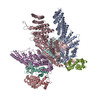
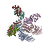
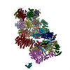
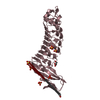
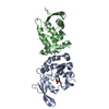
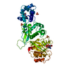
| Title | Formation of an intricate helical bundle dictates the assembly of the 26S proteasome lid | ||||||
 Components Components | (26S proteasome regulatory subunit ...) x 8 | ||||||
 Keywords Keywords | PROTEIN BINDING / alpha helix bundle / hybrid method / flexible fitting | ||||||
| Function / homology |  Function and homology information Function and homology informationMetalloprotease DUBs / peroxisome fission / proteasome storage granule assembly / protein deneddylation / COP9 signalosome / proteasome regulatory particle / proteasome regulatory particle, lid subcomplex / mitochondrial fission / metal-dependent deubiquitinase activity / Cross-presentation of soluble exogenous antigens (endosomes) ...Metalloprotease DUBs / peroxisome fission / proteasome storage granule assembly / protein deneddylation / COP9 signalosome / proteasome regulatory particle / proteasome regulatory particle, lid subcomplex / mitochondrial fission / metal-dependent deubiquitinase activity / Cross-presentation of soluble exogenous antigens (endosomes) / TNFR2 non-canonical NF-kB pathway / Ubiquitin Mediated Degradation of Phosphorylated Cdc25A / Regulation of PTEN stability and activity / KEAP1-NFE2L2 pathway / proteasome binding / CDK-mediated phosphorylation and removal of Cdc6 / Neddylation / FBXL7 down-regulates AURKA during mitotic entry and in early mitosis / regulation of protein catabolic process / Orc1 removal from chromatin / MAPK6/MAPK4 signaling / proteasome storage granule / Antigen processing: Ubiquitination & Proteasome degradation / proteasome assembly / Ub-specific processing proteases / enzyme regulator activity / Neutrophil degranulation / proteasome complex / metallopeptidase activity / ubiquitin-dependent protein catabolic process / proteasome-mediated ubiquitin-dependent protein catabolic process / ubiquitinyl hydrolase 1 / cysteine-type deubiquitinase activity / structural molecule activity / mitochondrion / nucleus / metal ion binding / cytoplasm / cytosol Similarity search - Function | ||||||
| Biological species |  | ||||||
| Method | ELECTRON MICROSCOPY / single particle reconstruction / cryo EM / Resolution: 7.4 Å | ||||||
 Authors Authors | Estrin, E. / Lopez-Blanco, J.R. / Chacon, P. / Martin, A. | ||||||
 Citation Citation | Journal: Proc Natl Acad Sci U S A / Year: 2012 Title: Near-atomic resolution structural model of the yeast 26S proteasome. Authors: Florian Beck / Pia Unverdorben / Stefan Bohn / Andreas Schweitzer / Günter Pfeifer / Eri Sakata / Stephan Nickell / Jürgen M Plitzko / Elizabeth Villa / Wolfgang Baumeister / Friedrich Förster /  Abstract: The 26S proteasome operates at the executive end of the ubiquitin-proteasome pathway. Here, we present a cryo-EM structure of the Saccharomyces cerevisiae 26S proteasome at a resolution of 7.4 Å or ...The 26S proteasome operates at the executive end of the ubiquitin-proteasome pathway. Here, we present a cryo-EM structure of the Saccharomyces cerevisiae 26S proteasome at a resolution of 7.4 Å or 6.7 Å (Fourier-Shell Correlation of 0.5 or 0.3, respectively). We used this map in conjunction with molecular dynamics-based flexible fitting to build a near-atomic resolution model of the holocomplex. The quality of the map allowed us to assign α-helices, the predominant secondary structure element of the regulatory particle subunits, throughout the entire map. We were able to determine the architecture of the Rpn8/Rpn11 heterodimer, which had hitherto remained elusive. The MPN domain of Rpn11 is positioned directly above the AAA-ATPase N-ring suggesting that Rpn11 deubiquitylates substrates immediately following commitment and prior to their unfolding by the AAA-ATPase module. The MPN domain of Rpn11 dimerizes with that of Rpn8 and the C-termini of both subunits form long helices, which are integral parts of a coiled-coil module. Together with the C-terminal helices of the six PCI-domain subunits they form a very large coiled-coil bundle, which appears to serve as a flexible anchoring device for all the lid subunits. | ||||||
| History |
| ||||||
| Remark 0 | THIS ENTRY 3J47 CONTAINS A STRUCTURAL MODEL FIT TO AN ELECTRON MICROSCOPY MAP (EMD-2165) DETERMINED ...THIS ENTRY 3J47 CONTAINS A STRUCTURAL MODEL FIT TO AN ELECTRON MICROSCOPY MAP (EMD-2165) DETERMINED ORIGINALLY BY AUTHORS: F.BECK, P.UNVERDORBEN, S.BOHN, A.SCHWEITZER, G.PFEIFER, E.SAKATA, S.NICKELL, J.M.PLITZKO, E.VILLA, W.BAUMEISTER, F.FORSTER |
- Structure visualization
| Movie |
Movie viewer |
|---|---|
| Structure viewer | Molecule: MolmilJmol/JSmol |
- Downloads & links
-Download
| PDBx/mmCIF format | 3j47.cif.gz | 68.9 KB | Display | PDBx/mmCIF format |
|---|---|---|---|---|
| PDB format | pdb3j47.ent.gz | 47.9 KB | Display | PDB format |
| PDBx/mmJSON format | 3j47.json.gz | Tree view | PDBx/mmJSON format | |
| Others |  Other downloads Other downloads |
-Validation report
| Summary document | 3j47_validation.pdf.gz | 705.2 KB | Display | wwPDB validaton report |
|---|---|---|---|---|
| Full document | 3j47_full_validation.pdf.gz | 767.1 KB | Display | |
| Data in XML | 3j47_validation.xml.gz | 28 KB | Display | |
| Data in CIF | 3j47_validation.cif.gz | 36.2 KB | Display | |
| Arichive directory | https://data.pdbj.org/pub/pdb/validation_reports/j4/3j47ftp://data.pdbj.org/pub/pdb/validation_reports/j4/3j47 | HTTPS FTP |
-Related structure data
| Related structure data |  2165M M: map data used to model this data |
|---|---|
| Similar structure data |


-Links
 PDBj
PDBj


- Assembly
| Deposited unit | 
|
|---|---|
| 1 |
|
-Components
-26S proteasome regulatory subunit ... , 8 types, 8 molecules VUOPQRST
| #1: Protein | Mass: 8013.857 Da / Num. of mol.: 1 Fragment: last three C-terminal helices (UNP residues 230-298) Source method: isolated from a natural source / Source: (natural) |
|---|---|
| #2: Protein | Mass: 13965.031 Da / Num. of mol.: 1 Fragment: last three C-terminal helices (UNP residues 188-308) Source method: isolated from a natural source / Source: (natural) |
| #3: Protein/peptide | Mass: 3353.889 Da / Num. of mol.: 1 / Fragment: C-terminal helix (UNP residues 360-387) / Source method: isolated from a natural source / Source: (natural) |
| #4: Protein/peptide | Mass: 3984.469 Da / Num. of mol.: 1 / Fragment: C-terminal helix (UNP residues 409-442) / Source method: isolated from a natural source / Source: (natural) |
| #5: Protein/peptide | Mass: 2741.054 Da / Num. of mol.: 1 / Fragment: C-terminal helix (UNP residues 407-431) / Source method: isolated from a natural source / Source: (natural) |
| #6: Protein/peptide | Mass: 2890.363 Da / Num. of mol.: 1 / Fragment: C-terminal helix (UNP residues 397-422) / Source method: isolated from a natural source / Source: (natural) |
| #7: Protein/peptide | Mass: 2924.156 Da / Num. of mol.: 1 / Fragment: C-terminal helix (UNP residues 455-478) / Source method: isolated from a natural source / Source: (natural) |
| #8: Protein/peptide | Mass: 1955.234 Da / Num. of mol.: 1 / Fragment: C-terminal helix (UNP residues 256-272) / Source method: isolated from a natural source / Source: (natural) |
-Experimental details
-Experiment
| Experiment | Method: ELECTRON MICROSCOPY |
|---|---|
| EM experiment | Aggregation state: PARTICLE / 3D reconstruction method: single particle reconstruction |
- Sample preparation
| Component | Name: 26S proteasome / Type: COMPLEX |
|---|---|
| Buffer solution | pH: 7.1 |
| Specimen | Embedding applied: NO / Shadowing applied: NO / Staining applied: NO / Vitrification applied: YES |
| Vitrification | Instrument: HOMEMADE PLUNGER / Cryogen name: ETHANE |
- Electron microscopy imaging
| Experimental equipment |  Model: Titan Krios / Image courtesy: FEI Company |
|---|---|
| Microscopy | Model: FEI TITAN KRIOS / Date: Mar 15, 2012 |
| Electron gun | Electron source:  FIELD EMISSION GUN / Accelerating voltage: 200 kV / Illumination mode: FLOOD BEAM FIELD EMISSION GUN / Accelerating voltage: 200 kV / Illumination mode: FLOOD BEAM |
| Electron lens | Mode: BRIGHT FIELD / Nominal magnification: 150000 X / Nominal defocus max: 3500 nm / Nominal defocus min: 1500 nm |
| Image recording | Electron dose: 25 e/Å2 / Film or detector model: TVIPS TEMCAM-F816 (8k x 8k) |
| Radiation | Protocol: SINGLE WAVELENGTH / Monochromatic (M) / Laue (L): M / Scattering type: x-ray |
| Radiation wavelength | Relative weight: 1 |
- Processing
| EM software |
| ||||||||||||
|---|---|---|---|---|---|---|---|---|---|---|---|---|---|
| Symmetry | Point symmetry: C1 (asymmetric) | ||||||||||||
| 3D reconstruction | Resolution: 7.4 Å / Resolution method: FSC 0.5 CUT-OFF / Num. of particles: 2464694 / Symmetry type: POINT | ||||||||||||
| Atomic model building | Protocol: FLEXIBLE FIT / Space: REAL / Target criteria: Cross-correlation coefficient Details: METHOD--Hybrid method + flexible fitting REFINEMENT PROTOCOL--Hybrid method DETAILS--Initial model was done with an in house hybrid method (EMTEGRATOR) that integrates topology constraints ...Details: METHOD--Hybrid method + flexible fitting REFINEMENT PROTOCOL--Hybrid method DETAILS--Initial model was done with an in house hybrid method (EMTEGRATOR) that integrates topology constraints with EM-map derived constraints. iMODFIT was then used for final flexible fitting. | ||||||||||||
| Refinement step | Cycle: LAST
|
