Movie
Movie Controller

[English] 日本語
 Yorodumi
- PDB-3j0e: Models for the T. thermophilus ribosome recycling factor and the ... -
+ Open data

- Basic information
| Entry | Database: PDB / ID: 3j0e | ||||||
|---|---|---|---|---|---|---|---|
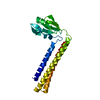
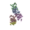
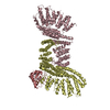
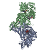
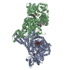
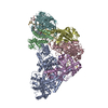
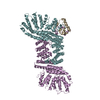
| Title | Models for the T. thermophilus ribosome recycling factor and the E. coli elongation factor G bound to the E. coli post-termination complex | ||||||
 Components Components |
| ||||||
 Keywords Keywords |  TRANSLATION / ribosome / ribosome recycling factor / Elongation Factor G TRANSLATION / ribosome / ribosome recycling factor / Elongation Factor G | ||||||
| Function / homology |  Function and homology information Function and homology informationribosome disassembly / guanosine tetraphosphate binding / translational elongation / misfolded RNA binding / Group I intron splicing / RNA folding / translational termination / translation elongation factor activity / positive regulation of RNA splicing / maintenance of translational fidelity ...ribosome disassembly / guanosine tetraphosphate binding / translational elongation / misfolded RNA binding / Group I intron splicing / RNA folding / translational termination / translation elongation factor activity / positive regulation of RNA splicing / maintenance of translational fidelity / Hydrolases; Acting on acid anhydrides; Acting on GTP to facilitate cellular and subcellular movement / cytosolic small ribosomal subunit / cytoplasmic translation / tRNA binding / rRNA binding / ribosome / structural constituent of ribosome / translation / response to antibiotic / GTPase activity / GTP binding / cytosol / cytoplasmSimilarity search - Function | ||||||
| Biological species |  Escherichia coli (E. coli) Escherichia coli (E. coli) Thermus thermophilus (bacteria) Thermus thermophilus (bacteria) | ||||||
| Method | ELECTRON MICROSCOPY / single particle reconstruction / cryo EM / Resolution: 9.9 Å | ||||||
 Authors Authors | Yokoyama, T. / Shaikh, T.R. / Iwakura, N. / Kaji, H. / Kaji, A. / Agrawal, R.K. | ||||||
 Citation Citation | Journal: EMBO J / Year: 2012 Title: Structural insights into initial and intermediate steps of the ribosome-recycling process. Authors: Takeshi Yokoyama / Tanvir R Shaikh / Nobuhiro Iwakura / Hideko Kaji / Akira Kaji / Rajendra K Agrawal /  Abstract: The ribosome-recycling factor (RRF) and elongation factor-G (EF-G) disassemble the 70S post-termination complex (PoTC) into mRNA, tRNA, and two ribosomal subunits. We have determined cryo-electron ...The ribosome-recycling factor (RRF) and elongation factor-G (EF-G) disassemble the 70S post-termination complex (PoTC) into mRNA, tRNA, and two ribosomal subunits. We have determined cryo-electron microscopic structures of the PoTC·RRF complex, with and without EF-G. We find that domain II of RRF initially interacts with universally conserved residues of the 23S rRNA helices 43 and 95, and protein L11 within the 50S ribosomal subunit. Upon EF-G binding, both RRF and tRNA are driven towards the tRNA-exit (E) site, with a large rotational movement of domain II of RRF towards the 30S ribosomal subunit. During this intermediate step of the recycling process, domain II of RRF and domain IV of EF-G adopt hitherto unknown conformations. Furthermore, binding of EF-G to the PoTC·RRF complex reverts the ribosome from ratcheted to unratcheted state. These results suggest that (i) the ribosomal intersubunit reorganizations upon RRF binding and subsequent EF-G binding could be instrumental in destabilizing the PoTC and (ii) the modes of action of EF-G during tRNA translocation and ribosome-recycling steps are markedly different. | ||||||
| History |
|
- Structure visualization
| Movie |
Movie viewer |
|---|---|
| Structure viewer | Molecule: MolmilJmol/JSmol |
- Downloads & links
-Download
| PDBx/mmCIF format | 3j0e.cif.gz | 236.7 KB | Display | PDBx/mmCIF format |
|---|---|---|---|---|
| PDB format | pdb3j0e.ent.gz | 179.6 KB | Display | PDB format |
| PDBx/mmJSON format | 3j0e.json.gz | Tree view | PDBx/mmJSON format | |
| Others |  Other downloads Other downloads |
-Validation report
| Arichive directory | https://data.pdbj.org/pub/pdb/validation_reports/j0/3j0eftp://data.pdbj.org/pub/pdb/validation_reports/j0/3j0e | HTTPS FTP |
|---|
-Related structure data
| Related structure data | 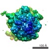 1917MC  1918MC 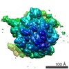 1915C 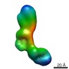 1916C 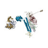 3j0dC M: map data used to model this data C: citing same article ( |
|---|---|
| Similar structure data |


-Links
 PDBj
PDBj


- Assembly
| Deposited unit | 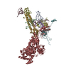
|
|---|---|
| 1 |
|
| Details | THE FULL BIOLOGICAL ASSEMBLY IS A COMPLEX OF MRNA, TRNA, RRF, EF-G, AND RIBOSOME. REMARK 350 REFERS ONLY TO INDIVIDUAL CHAINS THAT WERE MODELED AND DOES NOT REPRESENT THE FULL BIOLOGICAL ASSEMBLY. |
-Components
-Ribosomal 23S ... , 4 types, 4 molecules ABCD
| #1: RNA chain | Mass: 6994.202 Da / Num. of mol.: 1 / Fragment: helix 69 / Source method: isolated from a natural source / Source: (natural) Escherichia coli (E. coli) / References: GenBank: U00096.2 |
|---|---|
| #2: RNA chain | Mass: 5459.285 Da / Num. of mol.: 1 / Fragment: helix 71 / Source method: isolated from a natural source / Source: (natural) Escherichia coli (E. coli) / References: GenBank: U00096.2 |
| #3: RNA chain | Mass: 4228.565 Da / Num. of mol.: 1 / Fragment: helix 80 / Source method: isolated from a natural source / Source: (natural) Escherichia coli (E. coli) / References: GenBank: U00096.2 |
| #4: RNA chain | Mass: 6117.697 Da / Num. of mol.: 1 / Fragment: helix 93 / Source method: isolated from a natural source / Source: (natural) Escherichia coli (E. coli) / References: GenBank: U00096.2 |
-Ribosomal 16S ... , 2 types, 2 molecules Ee
| #5: RNA chain | Mass: 5787.508 Da / Num. of mol.: 1 / Fragment: helix 44 strand 1 / Source method: isolated from a natural source / Source: (natural) Escherichia coli (E. coli) / References: GenBank: U00096.2 |
|---|---|
| #6: RNA chain | Mass: 6173.722 Da / Num. of mol.: 1 / Fragment: helix 44 strand 2 / Source method: isolated from a natural source / Source: (natural) Escherichia coli (E. coli) / References: GenBank: U00096.2 |
-Protein , 3 types, 3 molecules FGH
| #7: Protein | Mass: 13636.961 Da / Num. of mol.: 1 / Source method: isolated from a natural source / Source: (natural) Escherichia coli (E. coli) / References: UniProt: P0A7S3 |
|---|---|
| #8: Protein | Mass: 21029.068 Da / Num. of mol.: 1 / Source method: isolated from a natural source / Source: (natural) Thermus thermophilus (bacteria) / References: UniProt: Q9WX76 |
| #9: Protein | EF-G / EF-G Mass: 77415.852 Da / Num. of mol.: 1 / Source method: isolated from a natural source / Source: (natural) Escherichia coli (E. coli) / References: UniProt: P0A6M8 |
-Details
| Sequence details | RIBOSOMAL RNA IS ONLY PARTIALLY MODELED IN THIS ENTRY: CHAINS A-D ARE PARTS OF THE 23S RIBOSOMAL ...RIBOSOMAL RNA IS ONLY PARTIALLY MODELED IN THIS ENTRY: CHAINS A-D ARE PARTS OF THE 23S RIBOSOMAL RNA AND CHAINS E AND e ARE PARTS OF THE 16S RIBOSOMAL RNA. |
|---|
-Experimental details
-Experiment
| Experiment | Method: ELECTRON MICROSCOPY |
|---|---|
| EM experiment | Aggregation state: PARTICLE / 3D reconstruction method: single particle reconstruction |
- Sample preparation
| Component | Name: E. COLI POST TERMINATION COMPLEX, T.THERMOPHILUS RIBOSOME RECYCLING FACTOR, E.COLI ELONGATION FACTOR G Type: RIBOSOME / Details: POTC-TTRRF-ECEFG-GDP-FUSIDIC A |
|---|---|
| Buffer solution | Name: BUFFER R / pH: 7.5 / Details: BUFFER R |
| Specimen | Conc.: 0.08 mg/ml / Embedding applied: NO / Shadowing applied: NO / Staining applied: NO / Vitrification applied: YES |
| Specimen support | Details: QUANTIFOIL HOLEY CARBON FILM G |
| Vitrification | Instrument: FEI VITROBOT MARK I / Cryogen name: ETHANE / Details: VITROBOT |
- Electron microscopy imaging
| Experimental equipment |  Model: Tecnai F20 / Image courtesy: FEI Company |
|---|---|
| Microscopy | Model: FEI TECNAI F20 / Date: Jan 22, 2010 |
| Electron gun | Electron source: FIELD EMISSION GUN / Accelerating voltage: 200 kV / Illumination mode: FLOOD BEAM |
| Electron lens | Mode: BRIGHT FIELDBright-field microscopy / Nominal magnification: 50000 X / Calibrated magnification: 50310 X / Nominal defocus max: 4000 nm / Nominal defocus min: 400 nm |
| Specimen holder | Temperature: 93 K / Tilt angle max: 0 ° / Tilt angle min: 0 ° |
| Image recording | Film or detector model: KODAK SO-163 FILM |
- Processing
| EM software |
| ||||||||||||||||||||||||||||
|---|---|---|---|---|---|---|---|---|---|---|---|---|---|---|---|---|---|---|---|---|---|---|---|---|---|---|---|---|---|
| CTF correction | Details: CTF CORRECTION OF 3D MAPS BY WIENER FILTRATION | ||||||||||||||||||||||||||||
| Symmetry | Point symmetry: C1 (asymmetric) | ||||||||||||||||||||||||||||
| 3D reconstruction | Method: 3D PROJECTION MATCHING / Resolution: 9.9 Å / Num. of particles: 338823 / Nominal pixel size: 2.78 Å / Symmetry type: POINT | ||||||||||||||||||||||||||||
| Atomic model building | Protocol: FLEXIBLE FIT / Details: REFINEMENT PROTOCOL--FLEXIBLE FITTING (MDFF) | ||||||||||||||||||||||||||||
| Atomic model building |
| ||||||||||||||||||||||||||||
| Refinement step | Cycle: LAST
|