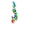
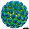
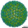
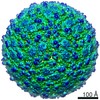
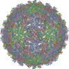
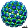
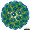
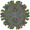
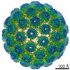
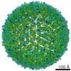
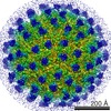
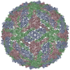
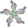
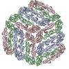
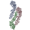
Journal: J Struct Biol / Year: 2013 Title: Membrane curvature in flaviviruses. Authors: Wei Zhang / Bärbel Kaufmann / Paul R Chipman / Richard J Kuhn / Michael G Rossmann / Abstract: Coordinated interplay between membrane proteins and the lipid bilayer is required for such processes as transporter function and the entrance of enveloped viruses into host cells. In this study, ...Coordinated interplay between membrane proteins and the lipid bilayer is required for such processes as transporter function and the entrance of enveloped viruses into host cells. In this study, three-dimensional cryo-electron microscopy density maps of mature and immature flaviviruses were analyzed to assess the curvature of the membrane leaflets and its relation to membrane-bound viral glycoproteins. The overall morphology of the viral membrane is determined by the icosahedral scaffold composed of envelope (E) and membrane (M) proteins through interaction of the proteins' stem-anchor regions with the membrane. In localized regions, small membrane areas exhibit convex, concave, flat or saddle-shaped surfaces that are constrained by the specific protein organization within each membrane leaflet. These results suggest that the organization of membrane proteins in small enveloped viruses mediate the formation of membrane curvature.
Name: TNE (12 mM Tris, 120 mM NaCl, 1 mM EDTA) / pH: 8 / Details: 12 mM Tris-HCL, 120 mM NaCl, 1 mM EDTA
Specimen
Conc.: 1 mg/ml / Embedding applied: NO / Shadowing applied: NO / Staining applied: NO / Vitrification applied: YES / Details: 12 mM Tris-HCL, 120 mM NaCl, 1 mM EDTA
Specimen support
Details: 400 mesh holey carbon copper grid
Vitrification
Instrument: HOMEMADE PLUNGER / Cryogen name: ETHANE / Temp: 98 K / Details: vitrification carried out in a BSL3 lab Method: A small vial of ethane is placed inside a larger liquid nitrogen reservoir. The grid holding a few microliters of the sample is held in place at the bottom of a plunger by means of fine ...Method: A small vial of ethane is placed inside a larger liquid nitrogen reservoir. The grid holding a few microliters of the sample is held in place at the bottom of a plunger by means of fine tweezers. Once the ethane in the vial is completely frozen, it needs to be melted slightly. When the liquid ethane is ready, a piece of filter paper is then pressed against the sample to blot off excess buffer sufficient to leave a thin layer on the grid. After a predetermined time, the filter paper is removed and the plunger is allowed to drop into the liquid ethane. Once the grid enters the liquid ethane, the sample is rapidly frozen and the grid is transferred under liquid nitrogen to a storage box immersed in liquid nitrogen for later use in the microscope.
-
Electron microscopy imaging
Microscopy
Model: FEI/PHILIPS CM300FEG/T / Date: Oct 21, 2004
Electron gun
Electron source: FIELD EMISSION GUN / Accelerating voltage: 300 kV / Illumination mode: FLOOD BEAM
Electron lens
Mode: BRIGHT FIELD / Calibrated magnification: 47440 X / Nominal defocus max: 321 nm / Nominal defocus min: 112 nm / Cs: 2 mm / Camera length: 0 mm
In the structure databanks used in Yorodumi, some data are registered as the other names, "COVID-19 virus" and "2019-nCoV". Here are the details of the virus and the list of structure data.
Jan 31, 2019. EMDB accession codes are about to change! (news from PDBe EMDB page)
EMDB accession codes are about to change! (news from PDBe EMDB page)
The allocation of 4 digits for EMDB accession codes will soon come to an end. Whilst these codes will remain in use, new EMDB accession codes will include an additional digit and will expand incrementally as the available range of codes is exhausted. The current 4-digit format prefixed with “EMD-” (i.e. EMD-XXXX) will advance to a 5-digit format (i.e. EMD-XXXXX), and so on. It is currently estimated that the 4-digit codes will be depleted around Spring 2019, at which point the 5-digit format will come into force.
The EM Navigator/Yorodumi systems omit the EMD- prefix.
Related info.:Q: What is EMD? / ID/Accession-code notation in Yorodumi/EM Navigator
Yorodumi is a browser for structure data from EMDB, PDB, SASBDB, etc.
This page is also the successor to EM Navigator detail page, and also detail information page/front-end page for Omokage search.
The word "yorodu" (or yorozu) is an old Japanese word meaning "ten thousand". "mi" (miru) is to see.
Related info.:EMDB / PDB / SASBDB / Comparison of 3 databanks / Yorodumi Search / Aug 31, 2016. New EM Navigator & Yorodumi / Yorodumi Papers / Jmol/JSmol / Function and homology information / Changes in new EM Navigator and Yorodumi
 Movie
Movie Controller


 Open data
 Basic information
 Components
 Keywords
 Function and homology information
 West Nile virus
 Authors
 Citation
 Structure visualization
 Downloads & links
 Other downloads
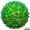
 PDBj
PDBj


 Assembly
 Sample preparation
 Electron microscopy imaging
 FIELD EMISSION GUN / Accelerating voltage: 300 kV / Illumination mode: FLOOD BEAM
 Processing