 Movie
Movie Controller

[English] 日本語
 Yorodumi
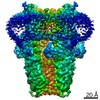
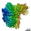
- PDB-3izo: Model of the fiber tail and its interactions with the penton base... -
+ Open data

- Basic information
| Entry | Database: PDB / ID: 3izo | ||||||
|---|---|---|---|---|---|---|---|
| Title | Model of the fiber tail and its interactions with the penton base of human adenovirus by cryo-electron microscopy | ||||||
 Components Components |
| ||||||
 Keywords Keywords |  VIRAL PROTEIN / human adenovirus fiber tail / pentameric penton base / trimeric fiber VIRAL PROTEIN / human adenovirus fiber tail / pentameric penton base / trimeric fiber | ||||||
| Function / homology |  Function and homology information Function and homology informationT=25 icosahedral viral capsid / adhesion receptor-mediated virion attachment to host cell / viral capsid / clathrin-dependent endocytosis of virus by host cell / cell adhesion / symbiont entry into host cell / host cell nucleus / structural molecule activity / virion attachment to host cell / nucleusSimilarity search - Function | ||||||
| Biological species |   Human adenovirus 5 Human adenovirus 5 | ||||||
| Method | ELECTRON MICROSCOPY / single particle reconstruction / cryo EM / Resolution: 3.6 Å | ||||||
 Authors Authors | Liu, H. | ||||||
 Citation Citation | Journal: J Mol Biol / Year: 2011 Title: Model of the trimeric fiber and its interactions with the pentameric penton base of human adenovirus by cryo-electron microscopy. Authors: Hongrong Liu / Lily Wu / Z Hong Zhou /  Abstract: Adenovirus invades host cells by first binding to host receptors through a trimeric fiber, which contains three domains: a receptor-binding knob domain, a long flexible shaft domain, and a penton ...Adenovirus invades host cells by first binding to host receptors through a trimeric fiber, which contains three domains: a receptor-binding knob domain, a long flexible shaft domain, and a penton base-attachment tail domain. Although the structure of the knob domain associated with a portion of the shaft has been solved by X-ray crystallography, the in situ structure of the fiber in the virion is not known; thus, it remains a mystery how the trimeric fiber attaches to its underlying pentameric penton base. By high-resolution cryo-electron microscopy, we have determined the structure of the human adenovirus type 5 (Ad5) to 3.6-Å resolution and have reported the full atomic models for its capsid proteins, but not for the fiber whose density cannot be directly interpreted due to symmetry mismatch with the penton base. Here, we report the determination of the Ad5 fiber structure and its mode of attachment to the pentameric penton base by using an integrative approach of multi-resolution filtering, homology modeling, computational simulation of mismatched symmetries, and fitting of atomic models into cryo-electron microscopy density maps. Our structure reveals that the interactions between the trimeric fiber and the pentameric penton base are mediated by a hydrophobic ring on the top surface of the penton base and three flexible tails inserted into three of the five available grooves formed by neighboring subunits of penton base. These interaction sites provide the molecular basis for the symmetry mismatch and can be targeted for optimizing adenovirus for gene therapy applications. | ||||||
| History |
|
- Structure visualization
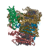
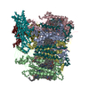
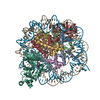
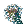
| Movie |
Movie viewer |
|---|---|
| Structure viewer | Molecule: MolmilJmol/JSmol |
- Downloads & links
-Download
| PDBx/mmCIF format | 3izo.cif.gz | 434.5 KB | Display | PDBx/mmCIF format |
|---|---|---|---|---|
| PDB format | pdb3izo.ent.gz | 327.6 KB | Display | PDB format |
| PDBx/mmJSON format | 3izo.json.gz | Tree view | PDBx/mmJSON format | |
| Others |  Other downloads Other downloads |
-Validation report
| Arichive directory | https://data.pdbj.org/pub/pdb/validation_reports/iz/3izoftp://data.pdbj.org/pub/pdb/validation_reports/iz/3izo | HTTPS FTP |
|---|
-Related structure data
| Related structure data | 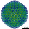 7034M M: map data used to model this data |
|---|---|
| Similar structure data |


-Links
 PDBj
PDBj

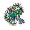
- Assembly
| Deposited unit | 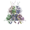
|
|---|---|
| 1 |
|
-Components
| #1: Protein | / Penton base protein / Virion component III / pIII Mass: 63356.602 Da / Num. of mol.: 5 / Source method: isolated from a natural source / Source: (natural) Human adenovirus 5 / Strain: HEK 293 cell / References: UniProt: P12538#2: Protein | Mass: 61633.066 Da / Num. of mol.: 3 / Source method: isolated from a natural source / Source: (natural) Human adenovirus 5 / Strain: HEK 293 cell / References: UniProt: Q7T416 |
|---|
-Experimental details
-Experiment
| Experiment | Method: ELECTRON MICROSCOPY |
|---|---|
| EM experiment | Aggregation state: PARTICLE / 3D reconstruction method: single particle reconstruction |
- Sample preparation
| Component | Name: Human adenovirus type 5 / Type: VIRUS |
|---|---|
| Buffer solution | pH: 7.5 / Details: 10mM Tris-HCL,1mM MgCl2 |
| Specimen | Embedding applied: NO / Shadowing applied: NO / Staining applied: NO / Vitrification applied: YES |
| Vitrification | Instrument: FEI VITROBOT MARK I / Cryogen name: ETHANE |
- Electron microscopy imaging
| Experimental equipment |  Model: Titan Krios / Image courtesy: FEI Company |
|---|---|
| Microscopy | Model: FEI TITAN KRIOS / Date: May 1, 2009 |
| Electron gun | Electron source: FIELD EMISSION GUN / Accelerating voltage: 200 kV / Illumination mode: FLOOD BEAM |
| Electron lens | Mode: BRIGHT FIELDBright-field microscopy / Nominal magnification: 59000 X / Nominal defocus max: 2500 nm / Nominal defocus min: 1000 nm |
| Image recording | Electron dose: 20 e/Å2 / Film or detector model: KODAK SO-163 FILM |
| Radiation | Protocol: SINGLE WAVELENGTH / Monochromatic (M) / Laue (L): M |
| Radiation wavelength | Relative weight: 1 |
- Processing
| EM software | Name: IMIRS / Category: 3D reconstruction | ||||||||||||
|---|---|---|---|---|---|---|---|---|---|---|---|---|---|
| Symmetry | Point symmetry: I (icosahedral) | ||||||||||||
| 3D reconstruction | Method: Cross-common lines for orientation and center refinement icosahedral symmetry-adapted functions for 3D reconstruction Resolution: 3.6 Å / Num. of particles: 31815 / Nominal pixel size: 1.076 Å / Actual pixel size: 1.1 Å / Symmetry type: POINT | ||||||||||||
| Refinement step | Cycle: LAST
|
