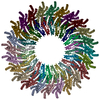
 Movie
Movie Controller

[English] 日本語
 Yorodumi
- PDB-3h3w: Fitting of the gp6 crystal structure into 3D cryo-EM reconstructi... -
+ Open data

- Basic information
| Entry | Database: PDB / ID: 3h3w | ||||||
|---|---|---|---|---|---|---|---|
| Title | Fitting of the gp6 crystal structure into 3D cryo-EM reconstruction of bacteriophage T4 dome-shaped baseplate | ||||||
 Components Components | Baseplate structural protein Gp6 | ||||||
 Keywords Keywords |  VIRAL PROTEIN / viral structural protein / Virion VIRAL PROTEIN / viral structural protein / Virion | ||||||
| Function / homology |  Function and homology information Function and homology information | ||||||
| Biological species |  Enterobacteria phage T4 (virus) Enterobacteria phage T4 (virus) | ||||||
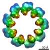
| Method | ELECTRON MICROSCOPY / single particle reconstruction / cryo EM / Resolution: 12 Å | ||||||
 Authors Authors | Aksyuk, A.A. / Leiman, P.G. / Shneider, M.M. / Mesyanzhinov, V.V. / Rossmann, M.G. | ||||||
 Citation Citation | Journal: Structure / Year: 2009 Title: The structure of gene product 6 of bacteriophage T4, the hinge-pin of the baseplate. Authors: Anastasia A Aksyuk / Petr G Leiman / Mikhail M Shneider / Vadim V Mesyanzhinov / Michael G Rossmann /  Abstract: The baseplate of bacteriophage T4 is a multicomponent protein complex, which controls phage attachment to the host. It assembles from six wedges and a central hub. During infection the baseplate ...The baseplate of bacteriophage T4 is a multicomponent protein complex, which controls phage attachment to the host. It assembles from six wedges and a central hub. During infection the baseplate undergoes a large conformational change from a dome-shaped to a flat, star-shaped structure. We report the crystal structure of the C-terminal half of gene product (gp) 6 and investigate its motion with respect to the other proteins during the baseplate rearrangement. Six gp6 dimers interdigitate, forming a ring that maintains the integrity of the baseplate in both conformations. One baseplate wedge contains an N-terminal dimer of gp6, whereas neighboring wedges are tied together through the C-terminal dimer of gp6. The dimeric interactions are preserved throughout the rearrangement of the baseplate. However, the hinge angle between the N- and C-terminal parts of gp6 changes by approximately 15 degrees , accounting for a 10 A radial increase in the diameter of the gp6 ring. | ||||||
| History |
|
- Structure visualization
| Movie |
Movie viewer |
|---|---|
| Structure viewer | Molecule: MolmilJmol/JSmol |
- Downloads & links
-Download
| PDBx/mmCIF format | 3h3w.cif.gz | 747 KB | Display | PDBx/mmCIF format |
|---|---|---|---|---|
| PDB format | pdb3h3w.ent.gz | 623.7 KB | Display | PDB format |
| PDBx/mmJSON format | 3h3w.json.gz | Tree view | PDBx/mmJSON format | |
| Others |  Other downloads Other downloads |
-Validation report
| Arichive directory | https://data.pdbj.org/pub/pdb/validation_reports/h3/3h3wftp://data.pdbj.org/pub/pdb/validation_reports/h3/3h3w | HTTPS FTP |
|---|
-Related structure data
| Related structure data | 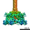 1048M 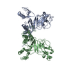 3h2tC 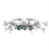 3h3yC C: citing same article ( M: map data used to model this data |
|---|---|
| Similar structure data |

-Links
 PDBj
PDBj- Assembly
| Deposited unit | 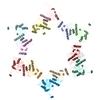
|
|---|---|
| 1 |
|
| Symmetry | Point symmetry: (Schoenflies symbol: C6 (6 fold cyclic)) |
-Components
| #1: Protein | Mass: 38304.449 Da / Num. of mol.: 12 / Fragment: gene product 6 deletion fragment / Mutation: residues 334-660 Source method: isolated from a genetically manipulated source Source: (gene. exp.) Enterobacteria phage T4 (virus) / Gene: 6 / Production host:  Escherichia coli (E. coli) / Strain (production host): BL21(DE3) / References: UniProt: P19060 Escherichia coli (E. coli) / Strain (production host): BL21(DE3) / References: UniProt: P19060 |
|---|
-Experimental details
-Experiment
| Experiment | Method: ELECTRON MICROSCOPY |
|---|---|
| EM experiment | Aggregation state: PARTICLE / 3D reconstruction method: single particle reconstruction |
- Sample preparation
| Component |
| |||||||||||||||
|---|---|---|---|---|---|---|---|---|---|---|---|---|---|---|---|---|
| Buffer solution | pH: 7 | |||||||||||||||
| Specimen | Conc.: 5 mg/ml / Embedding applied: NO / Shadowing applied: NO / Staining applied: NO / Vitrification applied: YES | |||||||||||||||
| Vitrification | Cryogen name: ETHANE |
- Electron microscopy imaging
| Microscopy | Model: FEI/PHILIPS CM300FEG/T / Date: Jan 30, 2001 |
|---|---|
| Electron gun | Electron source: FIELD EMISSION GUN / Accelerating voltage: 300 kV / Illumination mode: FLOOD BEAM |
| Electron lens | Mode: BRIGHT FIELDBright-field microscopy / Nominal magnification: 45000 X / Calibrated magnification: 47000 X / Nominal defocus max: 5000 nm / Nominal defocus min: 1200 nm |
| Specimen holder | Specimen holder model: GATAN LIQUID NITROGEN / Specimen holder type: 626 Single Tilt Cryotransfer System |
| Image recording | Electron dose: 25 e/Å2 / Film or detector model: KODAK SO-163 FILM |
| Radiation wavelength | Relative weight: 1 |
| Reflection | Highest resolution: 12 Å |
- Processing
| EM software |
| ||||||||||||
|---|---|---|---|---|---|---|---|---|---|---|---|---|---|
| Symmetry | Point symmetry: C6 (6 fold cyclic) | ||||||||||||
| 3D reconstruction | Resolution: 12 Å / Resolution method: FSC 0.5 CUT-OFF / Num. of particles: 945 / Symmetry type: POINT | ||||||||||||
| Atomic model building | Space: REAL | ||||||||||||
| Atomic model building | PDB-ID: 3H2T Accession code: 3H2T / Source name: PDB / Type: experimental model | ||||||||||||
| Refinement | Highest resolution: 12 Å | ||||||||||||
| Refinement step | Cycle: LAST / Highest resolution: 12 Å
|
