 Movie
Movie Controller

[English] 日本語
 Yorodumi
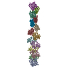
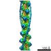
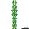
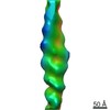
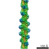
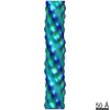
- PDB-3b5u: Actin filament model from extended form of acromsomal bundle in t... -
+ Open data

- Basic information
| Entry | Database: PDB / ID: 3b5u | ||||||
|---|---|---|---|---|---|---|---|
| Title | Actin filament model from extended form of acromsomal bundle in the Limulus sperm | ||||||
 Components Components | Actin, alpha skeletal muscle | ||||||
 Keywords Keywords | MOTOR PROTEIN / ACTIN FILAMENT / ACTIN / ACROMSOMAL BUNDLE / CRYOEM | ||||||
| Function / homology |  Function and homology information Function and homology informationcytoskeletal motor activator activity / tropomyosin binding / myosin heavy chain binding / mesenchyme migration / troponin I binding / actin filament bundle / filamentous actin / actin filament bundle assembly / skeletal muscle thin filament assembly / striated muscle thin filament ...cytoskeletal motor activator activity / tropomyosin binding / myosin heavy chain binding / mesenchyme migration / troponin I binding / actin filament bundle / filamentous actin / actin filament bundle assembly / skeletal muscle thin filament assembly / striated muscle thin filament / skeletal muscle myofibril / actin monomer binding / skeletal muscle fiber development / stress fiber / titin binding / actin filament polymerization / filopodium / actin filament / Hydrolases; Acting on acid anhydrides; Acting on acid anhydrides to facilitate cellular and subcellular movement / calcium-dependent protein binding / lamellipodium / cell body / hydrolase activity / protein domain specific binding / calcium ion binding / positive regulation of gene expression / magnesium ion binding / ATP binding / identical protein binding / cytoplasmSimilarity search - Function | ||||||
| Biological species |  Oryctolagus cuniculus (rabbit) Oryctolagus cuniculus (rabbit) | ||||||
| Method | ELECTRON CRYSTALLOGRAPHY / electron crystallography / cryo EM / Resolution: 9.5 Å | ||||||
 Authors Authors | Cong, Y. / Topf, M. / Sali, A. / Matsudaira, P. / Dougherty, M. / Chiu, W. / Schmid, M.F. | ||||||
 Citation Citation | Journal: J Mol Biol / Year: 2008 Title: Crystallographic conformers of actin in a biologically active bundle of filaments. Authors: Yao Cong / Maya Topf / Andrej Sali / Paul Matsudaira / Matthew Dougherty / Wah Chiu / Michael F Schmid /  Abstract: Actin carries out many of its cellular functions through its filamentous form; thus, understanding the detailed structure of actin filaments is an essential step in achieving a mechanistic ...Actin carries out many of its cellular functions through its filamentous form; thus, understanding the detailed structure of actin filaments is an essential step in achieving a mechanistic understanding of actin function. The acrosomal bundle in the Limulus sperm has been shown to be a quasi-crystalline array with an asymmetric unit composed of a filament with 14 actin-scruin pairs. The bundle in its true discharge state penetrates the jelly coat of the egg. Our previous electron crystallographic reconstruction demonstrated that the actin filament cross-linked by scruin in this acrosomal bundle state deviates significantly from a perfect F-actin helix. In that study, the tertiary structure of each of the 14 actin protomers in the asymmetric unit of the bundle filament was assumed to be constant. In the current study, an actin filament atomic model in the acrosomal bundle has been refined by combining rigid-body docking with multiple actin crystal structures from the Protein Data Bank and constrained energy minimization. Our observation demonstrates that actin protomers adopt different tertiary conformations when they form an actin filament in the bundle. The scruin and bundle packing forces appear to influence the tertiary and quaternary conformations of actin in the filament of this biologically active bundle. | ||||||
| History |
|
- Structure visualization
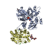
| Movie |
Movie viewer |
|---|---|
| Structure viewer | Molecule: MolmilJmol/JSmol |
- Downloads & links
-Download
| PDBx/mmCIF format | 3b5u.cif.gz | 812.5 KB | Display | PDBx/mmCIF format |
|---|---|---|---|---|
| PDB format | pdb3b5u.ent.gz | 644.7 KB | Display | PDB format |
| PDBx/mmJSON format | 3b5u.json.gz | Tree view | PDBx/mmJSON format | |
| Others |  Other downloads Other downloads |
-Validation report
| Arichive directory | https://data.pdbj.org/pub/pdb/validation_reports/b5/3b5uftp://data.pdbj.org/pub/pdb/validation_reports/b5/3b5u | HTTPS FTP |
|---|
-Related structure data
| Related structure data | 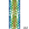 1088M  3b63C M: map data used to model this data C: citing same article ( |
|---|---|
| Similar structure data |
-Links
 PDBj
PDBj


- Assembly
| Deposited unit | 
|
|---|---|
| 1 |
|
-Components
| #1: Protein | / Alpha-actin-1 Mass: 42096.953 Da / Num. of mol.: 14 / Source method: isolated from a natural source Details: Holmes Actin model from Fiber Diffraction experiment Source: (natural) Oryctolagus cuniculus (rabbit) / References: UniProt: P68135 |
|---|
-Experimental details
-Experiment
| Experiment | Method: ELECTRON CRYSTALLOGRAPHY |
|---|---|
| EM experiment | Aggregation state: 3D ARRAY / 3D reconstruction method: electron crystallography |
- Sample preparation
| Component |
| |||||||||||||||
|---|---|---|---|---|---|---|---|---|---|---|---|---|---|---|---|---|
| Buffer solution | pH: 7.4 / Details: see J Mol Biol, 221, 711-725 (1991) | |||||||||||||||
| Specimen | Embedding applied: NO / Shadowing applied: NO / Staining applied: NO / Vitrification applied: YES | |||||||||||||||
| Vitrification | Instrument: HOMEMADE PLUNGER / Cryogen name: ETHANE |
-Data collection
| Microscopy | Model: JEOL 4000EX Details: For more information, please refer to EMDB entry EMD-1088 http://www.ebi.ac.uk/msd-srv/emsearch/atlas/1088_summary.html |
|---|---|
| Electron gun | Electron source: LAB6 / Accelerating voltage: 400 kV / Illumination mode: FLOOD BEAM |
| Electron lens | Mode: BRIGHT FIELDBright-field microscopy / Nominal magnification: 40000 X / Nominal defocus max: 3000 nm / Nominal defocus min: 800 nm |
| Specimen holder | Temperature: 106 K |
| Image recording | Electron dose: 15 e/Å2 / Film or detector model: KODAK SO-163 FILM |
- Processing
| EM software |
| ||||||||||||
|---|---|---|---|---|---|---|---|---|---|---|---|---|---|
| 3D reconstruction | Method: Cross-correlation and merging of crystallographic reflections derived from cryoelectron micrographs of 3D crystals. For more information, please refer to EMDB entry EMD-1088 http://www.ebi.ac. ...Method: Cross-correlation and merging of crystallographic reflections derived from cryoelectron micrographs of 3D crystals. For more information, please refer to EMDB entry EMD-1088 http://www.ebi.ac.uk/msd-srv/emsearch/atlas/1088_summary.html Resolution: 9.5 Å / Resolution method: DIFFRACTION PATTERN/LAYERLINES Details: Cross-correlation and merging of crystallographic reflections derived from cryoelectron micrographs of 3D crystalss: application to the Limulus acrosomal bundle, Michael F. Schmid, J. Struc. ...Details: Cross-correlation and merging of crystallographic reflections derived from cryoelectron micrographs of 3D crystalss: application to the Limulus acrosomal bundle, Michael F. Schmid, J. Struc. Biol., 144 (2003) 195-208 Symmetry type: 3D CRYSTAL | ||||||||||||
| Atomic model building | Protocol: AB INITIO MODEL / Space: REAL Target criteria: Best cross-correlation score between model and cryoEM map Details: REFINEMENT PROTOCOL--Foldhunter program from EMAN single particle analysis package | ||||||||||||
| Atomic model building | Details: Holmes' Actin model from Fiber Diffraction experiment | ||||||||||||
| Refinement step | Cycle: LAST
|
