Movie
Movie Controller

[English] 日本語
 Yorodumi
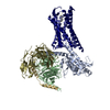
- EMDB-33889: Cryo-EM structure of the DC591053-bound human relaxin family pept... -
+ Open data

- Basic information
| Entry |  | ||||||||||||||||||||||||||||||||||||
|---|---|---|---|---|---|---|---|---|---|---|---|---|---|---|---|---|---|---|---|---|---|---|---|---|---|---|---|---|---|---|---|---|---|---|---|---|---|
| Title | Cryo-EM structure of the DC591053-bound human relaxin family peptide receptor 4 (RXFP4)-Gi complex | ||||||||||||||||||||||||||||||||||||
 Map data Map data | |||||||||||||||||||||||||||||||||||||
 Sample Sample |
| ||||||||||||||||||||||||||||||||||||
| Function / homology |  Function and homology information Function and homology informationRelaxin receptors / negative regulation of adenylate cyclase-activating adrenergic receptor signaling pathway involved in heart process / positive regulation of feeding behavior / G protein-coupled adenosine receptor signaling pathway / negative regulation of calcium ion-dependent exocytosis / positive regulation of urine volume / negative regulation of adenylate cyclase activity / positive regulation of neural precursor cell proliferation / G protein-coupled peptide receptor activity / gamma-aminobutyric acid signaling pathway ...Relaxin receptors / negative regulation of adenylate cyclase-activating adrenergic receptor signaling pathway involved in heart process / positive regulation of feeding behavior / G protein-coupled adenosine receptor signaling pathway / negative regulation of calcium ion-dependent exocytosis / positive regulation of urine volume / negative regulation of adenylate cyclase activity / positive regulation of neural precursor cell proliferation / G protein-coupled peptide receptor activity / gamma-aminobutyric acid signaling pathway / negative regulation of synaptic transmission / Activation of G protein gated Potassium channels / G-protein activation / G beta:gamma signalling through PI3Kgamma / Prostacyclin signalling through prostacyclin receptor / G beta:gamma signalling through PLC beta / ADP signalling through P2Y purinoceptor 1 / Thromboxane signalling through TP receptor / Presynaptic function of Kainate receptors / G beta:gamma signalling through CDC42 / Inhibition of voltage gated Ca2+ channels via Gbeta/gamma subunits / Glucagon-type ligand receptors / Adrenaline,noradrenaline inhibits insulin secretion / G alpha (12/13) signalling events / G beta:gamma signalling through BTK / ADP signalling through P2Y purinoceptor 12 / Cooperation of PDCL (PhLP1) and TRiC/CCT in G-protein beta folding / Thrombin signalling through proteinase activated receptors (PARs) / Ca2+ pathway / G alpha (z) signalling events / Extra-nuclear estrogen signaling / G alpha (s) signalling events / G alpha (q) signalling events / G alpha (i) signalling events / Glucagon-like Peptide-1 (GLP1) regulates insulin secretion / Vasopressin regulates renal water homeostasis via Aquaporins / neuronal dense core vesicle / regulation of calcium ion transport / negative regulation of apoptotic signaling pathway / neuropeptide signaling pathway / Adenylate cyclase inhibitory pathway / positive regulation of insulin receptor signaling pathway / positive regulation of vascular associated smooth muscle cell proliferation / adenylate cyclase-inhibiting G protein-coupled receptor signaling pathway / response to nutrient / positive regulation of superoxide anion generation / Regulation of insulin secretion / G protein-coupled receptor binding / Olfactory Signaling Pathway / G-protein beta/gamma-subunit complex binding / Activation of the phototransduction cascade / G beta:gamma signalling through PLC beta / Presynaptic function of Kainate receptors / Thromboxane signalling through TP receptor / G-protein activation / G protein-coupled acetylcholine receptor signaling pathway / Activation of G protein gated Potassium channels / Inhibition of voltage gated Ca2+ channels via Gbeta/gamma subunits / adenylate cyclase-activating G protein-coupled receptor signaling pathway / Prostacyclin signalling through prostacyclin receptor / Glucagon signaling in metabolic regulation / G beta:gamma signalling through CDC42 / ADP signalling through P2Y purinoceptor 12 / G beta:gamma signalling through BTK / Synthesis, secretion, and inactivation of Glucagon-like Peptide-1 (GLP-1) / Sensory perception of sweet, bitter, and umami (glutamate) taste / photoreceptor disc membrane / Adrenaline,noradrenaline inhibits insulin secretion / Glucagon-type ligand receptors / Vasopressin regulates renal water homeostasis via Aquaporins / G alpha (z) signalling events / cellular response to catecholamine stimulus / Glucagon-like Peptide-1 (GLP1) regulates insulin secretion / ADORA2B mediated anti-inflammatory cytokines production / ADP signalling through P2Y purinoceptor 1 / adenylate cyclase-activating dopamine receptor signaling pathway / G beta:gamma signalling through PI3Kgamma / cellular response to prostaglandin E stimulus / Cooperation of PDCL (PhLP1) and TRiC/CCT in G-protein beta folding / sensory perception of taste / GPER1 signaling / G-protein beta-subunit binding / Inactivation, recovery and regulation of the phototransduction cascade /  heterotrimeric G-protein complex / G alpha (12/13) signalling events / extracellular vesicle / signaling receptor complex adaptor activity / Thrombin signalling through proteinase activated receptors (PARs) / retina development in camera-type eye / GTPase binding / Ca2+ pathway / cell body / phospholipase C-activating G protein-coupled receptor signaling pathway / midbody / G alpha (i) signalling events / G alpha (s) signalling events / G alpha (q) signalling events / Ras protein signal transduction / cell population proliferation / Extra-nuclear estrogen signaling heterotrimeric G-protein complex / G alpha (12/13) signalling events / extracellular vesicle / signaling receptor complex adaptor activity / Thrombin signalling through proteinase activated receptors (PARs) / retina development in camera-type eye / GTPase binding / Ca2+ pathway / cell body / phospholipase C-activating G protein-coupled receptor signaling pathway / midbody / G alpha (i) signalling events / G alpha (s) signalling events / G alpha (q) signalling events / Ras protein signal transduction / cell population proliferation / Extra-nuclear estrogen signalingSimilarity search - Function | ||||||||||||||||||||||||||||||||||||
| Biological species |  Homo sapiens (human) / Homo sapiens (human) /  Bos taurus (cattle) / synthetic construct (others) Bos taurus (cattle) / synthetic construct (others) | ||||||||||||||||||||||||||||||||||||
| Method | single particle reconstruction / cryo EM / Resolution: 2.75 Å | ||||||||||||||||||||||||||||||||||||
 Authors Authors | Chen Y / Zhou QT / Wang J / Xu YW / Wang Y / Yan JH / Wang YB / Zhu Q / Zhao FH / Li CH ...Chen Y / Zhou QT / Wang J / Xu YW / Wang Y / Yan JH / Wang YB / Zhu Q / Zhao FH / Li CH / Chen CW / Cai XQ / Bathgate RAD / Shen C / Xu HE / Yang DH / Liu H / Wang MW | ||||||||||||||||||||||||||||||||||||
| Funding support |  China, 11 items China, 11 items
| ||||||||||||||||||||||||||||||||||||
 Citation Citation | Journal: Nat Commun / Year: 2023 Title: Ligand recognition mechanism of the human relaxin family peptide receptor 4 (RXFP4). Authors: Yan Chen / Qingtong Zhou / Jiang Wang / Youwei Xu / Yun Wang / Jiahui Yan / Yibing Wang / Qi Zhu / Fenghui Zhao / Chenghao Li / Chuan-Wei Chen / Xiaoqing Cai / Ross A D Bathgate / Chun Shen ...Authors: Yan Chen / Qingtong Zhou / Jiang Wang / Youwei Xu / Yun Wang / Jiahui Yan / Yibing Wang / Qi Zhu / Fenghui Zhao / Chenghao Li / Chuan-Wei Chen / Xiaoqing Cai / Ross A D Bathgate / Chun Shen / H Eric Xu / Dehua Yang / Hong Liu / Ming-Wei Wang /   Abstract: Members of the insulin superfamily regulate pleiotropic biological processes through two types of target-specific but structurally conserved peptides, insulin/insulin-like growth factors and ...Members of the insulin superfamily regulate pleiotropic biological processes through two types of target-specific but structurally conserved peptides, insulin/insulin-like growth factors and relaxin/insulin-like peptides. The latter bind to the human relaxin family peptide receptors (RXFPs). Here, we report three cryo-electron microscopy structures of RXFP4-G protein complexes in the presence of the endogenous ligand insulin-like peptide 5 (INSL5) or one of the two small molecule agonists, compound 4 and DC591053. The B chain of INSL5 adopts a single α-helix that penetrates into the orthosteric pocket, while the A chain sits above the orthosteric pocket, revealing a peptide-binding mode previously unknown. Together with mutagenesis and functional analyses, the key determinants responsible for the peptidomimetic agonism and subtype selectivity were identified. Our findings not only provide insights into ligand recognition and subtype selectivity among class A G protein-coupled receptors, but also expand the knowledge of signaling mechanisms in the insulin superfamily. | ||||||||||||||||||||||||||||||||||||
| History |
|
- Structure visualization
| Supplemental images |
|---|

- Downloads & links
-EMDB archive
| Map data | emd_33889.map.gz | 56.9 MB | EMDB map data format | |
|---|---|---|---|---|
| Header (meta data) | emd-33889-v30.xmlemd-33889.xml | 23.7 KB 23.7 KB | Display Display | EMDB header |
| FSC (resolution estimation) | emd_33889_fsc.xml | 9.4 KB | Display | FSC data file |
| Images |  emd_33889.png emd_33889.png | 104.5 KB | ||
| Others | emd_33889_half_map_1.map.gzemd_33889_half_map_2.map.gz | 59.5 MB 59.5 MB | ||
| Archive directory |  http://ftp.pdbj.org/pub/emdb/structures/EMD-33889ftp://ftp.pdbj.org/pub/emdb/structures/EMD-33889 http://ftp.pdbj.org/pub/emdb/structures/EMD-33889ftp://ftp.pdbj.org/pub/emdb/structures/EMD-33889 | HTTPS FTP |
-Related structure data
| Related structure data |  7yk7MC  7yj4C  7yk6C M: atomic model generated by this map C: citing same article ( |
|---|---|
| Similar structure data |


-Links
| EMDB pages | EMDB (EBI/PDBe) / EMDataResource |
|---|---|
| Related items in Molecule of the Month |


-Map
| File | Download / File: emd_33889.map.gz / Format: CCP4 / Size: 64 MB / Type: IMAGE STORED AS FLOATING POINT NUMBER (4 BYTES) | ||||||||||||||||||||
|---|---|---|---|---|---|---|---|---|---|---|---|---|---|---|---|---|---|---|---|---|---|
| Voxel size | X=Y=Z: 1.071 Å | ||||||||||||||||||||
| Density |
| ||||||||||||||||||||
| Symmetry | Space group: 1 | ||||||||||||||||||||
| Details | EMDB XML:
|
-Supplemental data
-Half map: #2
| File | emd_33889_half_map_1.map | ||||||||||||
|---|---|---|---|---|---|---|---|---|---|---|---|---|---|
| Projections & Slices |
| ||||||||||||
| Density Histograms |
 Z
Z Y
Y X
X


-Half map: #1
| File | emd_33889_half_map_2.map | ||||||||||||
|---|---|---|---|---|---|---|---|---|---|---|---|---|---|
| Projections & Slices |
| ||||||||||||
| Density Histograms |


- Sample components
+Entire : Cryo-EM structure of the human relaxin family peptide receptor 4 ...
+Supramolecule #1: Cryo-EM structure of the human relaxin family peptide receptor 4 ...
+Supramolecule #2: relaxin family peptide receptor 4
+Supramolecule #3: G protein
+Supramolecule #4: scFv16
+Macromolecule #1: Guanine nucleotide-binding protein G(I)/G(S)/G(O) subunit gamma-2


+Macromolecule #2: Guanine nucleotide-binding protein G(i) subunit alpha-2
+Macromolecule #3: Relaxin-3 receptor 2
+Macromolecule #4: scFv16
+Macromolecule #5: Guanine nucleotide-binding protein G(I)/G(S)/G(T) subunit beta-1
+Macromolecule #6: [(1S)-7-ethoxy-6-methoxy-1-[2-(5-methoxy-1H-indol-3-yl)ethyl]-3,4...

-Experimental details
-Structure determination
| Method | cryo EM |
|---|---|
 Processing Processing | single particle reconstruction |
| Aggregation state | particle |
-Sample preparation
| Buffer | pH: 7.4 |
|---|---|
| Vitrification | Cryogen name: ETHANE |
- Electron microscopy
| Microscope | FEI TITAN KRIOS |
|---|---|
| Electron beam | Acceleration voltage: 300 kV / Electron source: OTHER |
| Electron optics | Illumination mode: OTHER / Imaging mode: BRIGHT FIELDBright-field microscopy / Nominal defocus max: 2.2 µm / Nominal defocus min: 1.2 µm |
| Image recording | Film or detector model: GATAN K3 (6k x 4k) / Average electron dose: 80.0 e/Å2 |
| Experimental equipment |  Model: Titan Krios / Image courtesy: FEI Company |
-Image processing
| Startup model | Type of model: PDB ENTRY PDB model - PDB ID: Details: and 6D9H |
|---|---|
| Initial angle assignment | Type: OTHER |
| Final angle assignment | Type: OTHER |
| Final reconstruction | Resolution.type: BY AUTHOR / Resolution: 2.75 Å / Resolution method: DIFFRACTION PATTERN/LAYERLINES / Number images used: 225327 |
| FSC plot (resolution estimation) |  |