 Movie
Movie Controller

[English] 日本語
 Yorodumi
- EMDB-3282: Negative stain electron microscopy structure of TssA from EAEC ty... -
+ Open data

- Basic information
| Entry | Database: EMDB / ID: EMD-3282 | |||||||||
|---|---|---|---|---|---|---|---|---|---|---|
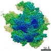
| Title | Negative stain electron microscopy structure of TssA from EAEC type VI secretion system | |||||||||
 Map data Map data | 3D Reconstruction of TssA dodecamer | |||||||||
 Sample Sample |
| |||||||||
 Keywords Keywords |  Type VI protein secretion system / bacterial contractile tail structure Type VI protein secretion system / bacterial contractile tail structure | |||||||||
| Biological species |  Escherichia coli 042 (bacteria) Escherichia coli 042 (bacteria) | |||||||||
| Method | single particle reconstruction / negative staining / Resolution: 19.0 Å | |||||||||
 Authors Authors | Durand E / Fronzes R / Cambillau C / Cascales E | |||||||||
 Citation Citation | Journal: Nature / Year: 2016 Title: Priming and polymerization of a bacterial contractile tail structure. Authors: Abdelrahim Zoued / Eric Durand / Yannick R Brunet / Silvia Spinelli / Badreddine Douzi / Mathilde Guzzo / Nicolas Flaugnatti / Pierre Legrand / Laure Journet / Rémi Fronzes / Tâm Mignot / ...Authors: Abdelrahim Zoued / Eric Durand / Yannick R Brunet / Silvia Spinelli / Badreddine Douzi / Mathilde Guzzo / Nicolas Flaugnatti / Pierre Legrand / Laure Journet / Rémi Fronzes / Tâm Mignot / Christian Cambillau / Eric Cascales /  Abstract: Contractile tails are composed of an inner tube wrapped by an outer sheath assembled in an extended, metastable conformation that stores mechanical energy necessary for its contraction. Contraction ...Contractile tails are composed of an inner tube wrapped by an outer sheath assembled in an extended, metastable conformation that stores mechanical energy necessary for its contraction. Contraction is used to propel the rigid inner tube towards target cells for DNA or toxin delivery. Although recent studies have revealed the structure of the contractile sheath of the type VI secretion system, the mechanisms by which its polymerization is controlled and coordinated with the assembly of the inner tube remain unknown. Here we show that the starfish-like TssA dodecameric complex interacts with tube and sheath components. Fluorescence microscopy experiments in enteroaggregative Escherichia coli reveal that TssA binds first to the type VI secretion system membrane core complex and then initiates tail polymerization. TssA remains at the tip of the growing structure and incorporates new tube and sheath blocks. On the basis of these results, we propose that TssA primes and coordinates tail tube and sheath biogenesis. | |||||||||
| History |
|
- Structure visualization
| Movie |
 Movie viewer Movie viewer |
|---|---|
| Structure viewer | EM map: SurfViewMolmilJmol/JSmol |
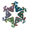
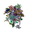
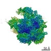
| Supplemental images |

- Downloads & links
-EMDB archive
| Map data | emd_3282.map.gz | 5.4 MB | EMDB map data format | |
|---|---|---|---|---|
| Header (meta data) | emd-3282-v30.xmlemd-3282.xml | 10.9 KB 10.9 KB | Display Display | EMDB header |
| Images | 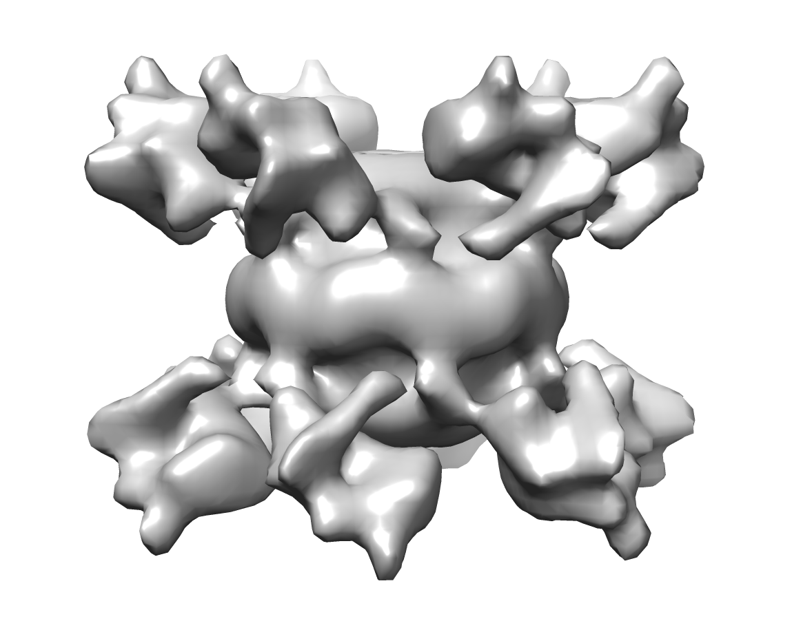 EMD-3282_TssA.png EMD-3282_TssA.png | 226.4 KB | ||
| Archive directory |  http://ftp.pdbj.org/pub/emdb/structures/EMD-3282ftp://ftp.pdbj.org/pub/emdb/structures/EMD-3282 http://ftp.pdbj.org/pub/emdb/structures/EMD-3282ftp://ftp.pdbj.org/pub/emdb/structures/EMD-3282 | HTTPS FTP |
-Related structure data


-Links
| EMDB pages | EMDB (EBI/PDBe) / EMDataResource |
|---|
-Map
| File | Download / File: emd_3282.map.gz / Format: CCP4 / Size: 29.8 MB / Type: IMAGE STORED AS FLOATING POINT NUMBER (4 BYTES) | ||||||||||||||||||||||||||||||||||||||||||||||||||||||||||||||||||||
|---|---|---|---|---|---|---|---|---|---|---|---|---|---|---|---|---|---|---|---|---|---|---|---|---|---|---|---|---|---|---|---|---|---|---|---|---|---|---|---|---|---|---|---|---|---|---|---|---|---|---|---|---|---|---|---|---|---|---|---|---|---|---|---|---|---|---|---|---|---|
| Annotation | 3D Reconstruction of TssA dodecamer | ||||||||||||||||||||||||||||||||||||||||||||||||||||||||||||||||||||
| Voxel size | X=Y=Z: 1.9 Å | ||||||||||||||||||||||||||||||||||||||||||||||||||||||||||||||||||||
| Density |
| ||||||||||||||||||||||||||||||||||||||||||||||||||||||||||||||||||||
| Symmetry | Space group: 1 | ||||||||||||||||||||||||||||||||||||||||||||||||||||||||||||||||||||
| Details | EMDB XML:
CCP4 map header:
| ||||||||||||||||||||||||||||||||||||||||||||||||||||||||||||||||||||
-Supplemental data
- Sample components
-Entire : TssA dodecamer
| Entire | Name: TssA dodecamer |
|---|---|
| Components |
|
-Supramolecule #1000: TssA dodecamer
| Supramolecule | Name: TssA dodecamer / type: sample / ID: 1000 / Details: The sample was mono disperse / Oligomeric state: 12 / Number unique components: 1 |
|---|---|
| Molecular weight | Experimental: 952 MDa / Theoretical: 891 MDa Method: on-line multi-angle laser light scattering/quasi-elastic light scattering/absorbance/refractive index (MALS/QELS/UV/RI) |
-Macromolecule #1: TssA
| Macromolecule | Name: TssA / type: protein_or_peptide / ID: 1 / Name.synonym: TssA / Number of copies: 12 / Oligomeric state: Dodecamer / Recombinant expression: Yes |
|---|---|
| Source (natural) | Organism: Escherichia coli 042 (bacteria) / Strain: strain 042 / synonym: Enteroaggregative E. coli / Location in cell: Cytoplasm |
| Molecular weight | Experimental: 74 KDa / Theoretical: 74 KDa |
| Recombinant expression | Organism: Escherichia coli (E. coli) / Recombinant strain: Escherichia coli BL21 / Recombinant plasmid: pIBA3C |
-Experimental details
-Structure determination
| Method | negative staining |
|---|---|
 Processing Processing | single particle reconstruction |
| Aggregation state | particle |
-Sample preparation
| Concentration | 0.01 mg/mL |
|---|---|
| Buffer | pH: 8 / Details: 20mM Tris-HCL, 150mM NaCl |
| Staining | Type: NEGATIVE Details: TssA sample was applied to glow-discharged carbon-coated copper grids (Agar Scientific). After 30 sec of absorption, the sample was blotted, washed with three drops of water and then stained ...Details: TssA sample was applied to glow-discharged carbon-coated copper grids (Agar Scientific). After 30 sec of absorption, the sample was blotted, washed with three drops of water and then stained with 2% uranyl acetate |
| Grid | Details: 400 mesh glow-discharged carbon-coated copper grids (Agar Scientific) |
| Vitrification | Cryogen name: NONE / Instrument: OTHER |
- Electron microscopy
| Microscope | FEI TECNAI F20 |
|---|---|
| Electron beam | Acceleration voltage: 200 kV / Electron source: FIELD EMISSION GUN |
| Electron optics | Illumination mode: SPOT SCAN / Imaging mode: BRIGHT FIELDBright-field microscopy / Cs: 2.10 mm / Nominal defocus max: 2.5 µm / Nominal defocus min: 0.5 µm / Nominal magnification: 50000 |
| Specialist optics | Energy filter - Name: FEI |
| Sample stage | Specimen holder model: OTHER |
| Alignment procedure | Legacy - Astigmatism: Objective lens astigmatism was corrected at 100,000 times magnification |
| Date | Jun 10, 2015 |
| Image recording | Category: CCD / Film or detector model: FEI FALCON II (4k x 4k) / Number real images: 500 / Average electron dose: 14 e/Å2 |
| Experimental equipment |  Model: Tecnai F20 / Image courtesy: FEI Company |
-Image processing
| CTF correction | Details: Each particle |
|---|---|
| Final two d classification | Number classes: 10 |
| Final reconstruction | Applied symmetry - Point group: D6 (2x6 fold dihedral) / Algorithm: OTHER / Resolution.type: BY AUTHOR / Resolution: 19.0 Å / Resolution method: OTHER / Software - Name: EMAN2, RELION / Number images used: 18000 |
| Details | A total of 100000 particles were automatically selected from 500 independent images and extracted within boxes of 180 pixels by 180 pixels using EMAN2/BOXER. The CTF was estimated and corrected by phase flipping using EMAN2. All two- and three-dimensional classifications and refinements were performed using RELION 1.3. We used three rounds of reference-free 2D class averaging to clean up the automatically selected dataset. Only highly populated classes displaying high-resolution features were conserved during this procedure and a final dataset of 20000 particles was assembled. An initial 3D-model was generated in EMAN2 using using 30 classes. 3D classification was then performed in Relion with 5 classes. The particles corresponding to most populated class were used for refinement. Relion auto-refine procedure was used to obtain a final reconstruction at 19A resolution after masking and with D6 symmetry imposed. Reported resolutions are based on the gold-standard Fourier shell correlation 0.143 criterion, and FSC curve were corrected for the effects of a soft mask on the FSC curve using high-resolution noise substitution. |
