 Movie
Movie Controller


+ Open data

- Basic information
| Entry | Database: EMDB / ID: EMD-3233 | |||||||||
|---|---|---|---|---|---|---|---|---|---|---|
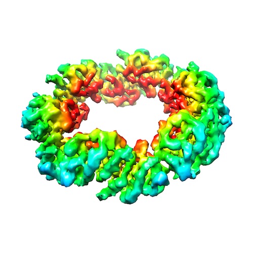
| Title | Volta phase plate cryo-EM of the small protein complex Prx3 | |||||||||
 Map data Map data | Volta phase plate reconstruction of hPrx3 | |||||||||
 Sample Sample |
| |||||||||
 Keywords Keywords | Peroxidase / antioxidant | |||||||||
| Function / homology |  Function and homology information Function and homology information | |||||||||
| Biological species |  Homo sapiens (human) Homo sapiens (human) | |||||||||
| Method | single particle reconstruction / cryo EM / Resolution: 4.4 Å | |||||||||
 Authors Authors | Khoshouei M / Radjainia M / Phillips AJ / Gerrard JA / Mitra AK / Plitzko JM / Baumeister W / Danev R | |||||||||
 Citation Citation | Journal: Nat Commun / Year: 2016 Title: Volta phase plate cryo-EM of the small protein complex Prx3. Authors: Maryam Khoshouei / Mazdak Radjainia / Amy J Phillips / Juliet A Gerrard / Alok K Mitra / Jürgen M Plitzko / Wolfgang Baumeister / Radostin Danev /    Abstract: Cryo-EM of large, macromolecular assemblies has seen a significant increase in the numbers of high-resolution structures since the arrival of direct electron detectors. However, sub-nanometre ...Cryo-EM of large, macromolecular assemblies has seen a significant increase in the numbers of high-resolution structures since the arrival of direct electron detectors. However, sub-nanometre resolution cryo-EM structures are rare compared with crystal structure depositions, particularly for relatively small particles (<400 kDa). Here we demonstrate the benefits of Volta phase plates for single-particle analysis by time-efficient cryo-EM structure determination of 257 kDa human peroxiredoxin-3 dodecamers at 4.4 Å resolution. The Volta phase plate improves the applicability of cryo-EM for small molecules and accelerates structure determination. | |||||||||
| History |
|
- Structure visualization
| Movie |
Movie viewer |
|---|---|
| Structure viewer | EM map: SurfViewMolmilJmol/JSmol |
| Supplemental images |
- Downloads & links
-EMDB archive
| Map data | emd_3233.map.gz | 1.3 MB | EMDB map data format | |
|---|---|---|---|---|
| Header (meta data) | emd-3233-v30.xmlemd-3233.xml | 8.8 KB 8.8 KB | Display Display | EMDB header |
| FSC (resolution estimation) | emd_3233_fsc.xml | 5.4 KB | Display | FSC data file |
| Images | EMD-3233.tif | 260.6 KB | ||
| Archive directory |  http://ftp.pdbj.org/pub/emdb/structures/EMD-3233ftp://ftp.pdbj.org/pub/emdb/structures/EMD-3233 http://ftp.pdbj.org/pub/emdb/structures/EMD-3233ftp://ftp.pdbj.org/pub/emdb/structures/EMD-3233 | HTTPS FTP |
-Validation report
| Summary document | emd_3233_validation.pdf.gz | 240.3 KB | Display | EMDB validaton report |
|---|---|---|---|---|
| Full document | emd_3233_full_validation.pdf.gz | 239.4 KB | Display | |
| Data in XML | emd_3233_validation.xml.gz | 8.5 KB | Display | |
| Arichive directory | https://ftp.pdbj.org/pub/emdb/validation_reports/EMD-3233ftp://ftp.pdbj.org/pub/emdb/validation_reports/EMD-3233 | HTTPS FTP |
-Related structure data
| Similar structure data | |
|---|---|
| EM raw data | EMPIAR-10050 (Title: Volta phase plate cryo-EM of the small protein complex Prx3 Data size: 612.5 / Data #1: VPP_Prx3_4.4res [micrographs - multiframe] / Data #2: VPP_Prx3_7.3res [micrographs - multiframe] / Data #3: No_VPP_mrc_files [micrographs - single frame] / Data #4: No_VPP_dat_files [micrographs - multiframe]) |


-Links
| EMDB pages | EMDB (EBI/PDBe) / EMDataResource |
|---|
-Map
| File | Download / File: emd_3233.map.gz / Format: CCP4 / Size: 13.1 MB / Type: IMAGE STORED AS FLOATING POINT NUMBER (4 BYTES) | ||||||||||||||||||||||||||||||||||||||||||||||||||||||||||||
|---|---|---|---|---|---|---|---|---|---|---|---|---|---|---|---|---|---|---|---|---|---|---|---|---|---|---|---|---|---|---|---|---|---|---|---|---|---|---|---|---|---|---|---|---|---|---|---|---|---|---|---|---|---|---|---|---|---|---|---|---|---|
| Annotation | Volta phase plate reconstruction of hPrx3 | ||||||||||||||||||||||||||||||||||||||||||||||||||||||||||||
| Voxel size | X=Y=Z: 1.74 Å | ||||||||||||||||||||||||||||||||||||||||||||||||||||||||||||
| Density |
| ||||||||||||||||||||||||||||||||||||||||||||||||||||||||||||
| Symmetry | Space group: 1 | ||||||||||||||||||||||||||||||||||||||||||||||||||||||||||||
| Details | EMDB XML:
CCP4 map header:
| ||||||||||||||||||||||||||||||||||||||||||||||||||||||||||||
-Supplemental data
- Sample components
-Entire : Human peroxiredoxin 3
| Entire | Name: Human peroxiredoxin 3 |
|---|---|
| Components |
|
-Supramolecule #1000: Human peroxiredoxin 3
| Supramolecule | Name: Human peroxiredoxin 3 / type: sample / ID: 1000 / Details: The sample was monodisperse / Oligomeric state: Dodecamer / Number unique components: 1 |
|---|---|
| Molecular weight | Theoretical: 257 KDa |
-Macromolecule #1: Peroxiredoxin 3
| Macromolecule | Name: Peroxiredoxin 3 / type: protein_or_peptide / ID: 1 / Number of copies: 12 / Oligomeric state: Dodecamer / Recombinant expression: Yes |
|---|---|
| Source (natural) | Organism: Homo sapiens (human) / synonym: Human / Organelle: Mitochondria / Location in cell: Matrix |
| Molecular weight | Theoretical: 257 KDa |
| Recombinant expression | Organism:  |
| Sequence | UniProtKB: Peroxiredoxin 3 isoform a variant |
-Experimental details
-Structure determination
| Method | cryo EM |
|---|---|
 Processing Processing | single particle reconstruction |
| Aggregation state | particle |
-Sample preparation
| Concentration | 0.5 mg/mL |
|---|---|
| Buffer | pH: 8 / Details: 150mM NaCl, 20mM Hepes |
| Grid | Details: Quantifoil R1.2/1.3 |
| Vitrification | Cryogen name: ETHANE-PROPANE MIXTURE / Chamber humidity: 85 % / Chamber temperature: 93 K / Instrument: FEI VITROBOT MARK III / Method: Blotting time: 4seconds |
- Electron microscopy
| Microscope | FEI TITAN KRIOS |
|---|---|
| Specialist optics | Energy filter - Name: Quantum post-column energy filter / Energy filter - Lower energy threshold: 0.0 eV / Energy filter - Upper energy threshold: 20.0 eV |
| Details | Volta Phase Plate |
| Date | Jul 1, 2015 |
| Image recording | Category: CCD / Film or detector model: GATAN K2 SUMMIT (4k x 4k) / Average electron dose: 20 e/Å2 |
| Electron beam | Acceleration voltage: 300 kV / Electron source:  FIELD EMISSION GUN FIELD EMISSION GUN |
| Electron optics | Calibrated magnification: 28700 / Illumination mode: FLOOD BEAM / Imaging mode: BRIGHT FIELD / Cs: 2.7 mm |
| Sample stage | Specimen holder model: FEI TITAN KRIOS AUTOGRID HOLDER |
| Experimental equipment |  Model: Titan Krios / Image courtesy: FEI Company |
-Image processing
| Final reconstruction | Applied symmetry - Point group: D6 (2x6 fold dihedral) / Resolution.type: BY AUTHOR / Resolution: 4.4 Å / Resolution method: OTHER / Software - Name: Relion / Number images used: 8000 |
|---|---|
| FSC plot (resolution estimation) |  |
