 Movie
Movie Controller

[English] 日本語
 Yorodumi
- EMDB-3231: Structure and function based design of Plasmodium-selective prote... -
+ Open data

- Basic information
| Entry | Database: EMDB / ID: EMD-3231 | |||||||||
|---|---|---|---|---|---|---|---|---|---|---|
| Title | Structure and function based design of Plasmodium-selective proteasome inhibitors | |||||||||
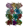
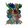
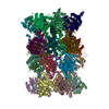
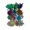
 Map data Map data | unsharpened and unfiltered 3D reconstruction of the Plasmodium falciparum 20S proteasome core with a ligand | |||||||||
 Sample Sample |
| |||||||||
 Keywords Keywords |  proteasome / 20S / Plasmodium / malaria / inhibitor / drug design / cryo-EM proteasome / 20S / Plasmodium / malaria / inhibitor / drug design / cryo-EM | |||||||||
| Function / homology |  Function and homology information Function and homology informationAUF1 (hnRNP D0) binds and destabilizes mRNA / Cross-presentation of soluble exogenous antigens (endosomes) / ABC-family proteins mediated transport / MAPK6/MAPK4 signaling / : / Orc1 removal from chromatin / CDK-mediated phosphorylation and removal of Cdc6 / KEAP1-NFE2L2 pathway / UCH proteinases / Ub-specific processing proteases ...AUF1 (hnRNP D0) binds and destabilizes mRNA / Cross-presentation of soluble exogenous antigens (endosomes) / ABC-family proteins mediated transport / MAPK6/MAPK4 signaling / : / Orc1 removal from chromatin / CDK-mediated phosphorylation and removal of Cdc6 / KEAP1-NFE2L2 pathway / UCH proteinases / Ub-specific processing proteases / Neddylation / Antigen processing: Ubiquitination & Proteasome degradation / Neutrophil degranulation / proteasome core complex / proteasomal ubiquitin-independent protein catabolic process / proteasome endopeptidase complex / proteasome core complex, beta-subunit complex / proteasome core complex, alpha-subunit complex / threonine-type endopeptidase activity / proteasomal protein catabolic process / ubiquitin-dependent protein catabolic process / proteasome-mediated ubiquitin-dependent protein catabolic process / endopeptidase activity / hydrolase activity / nucleus / cytosol / cytoplasmSimilarity search - Function | |||||||||
| Biological species |  Plasmodium falciparum (malaria parasite P. falciparum) / synthetic construct (others) Plasmodium falciparum (malaria parasite P. falciparum) / synthetic construct (others) | |||||||||
| Method | single particle reconstruction / cryo EM / Resolution: 3.6 Å | |||||||||
 Authors Authors | Li H / O' Donoghue AJ / van der Linden WA / Xie SC / Yoo E / Foe IT / Tilley L / Craik CS / da Fonseca PCA / Bogyo M | |||||||||
 Citation Citation | Journal: Nature / Year: 2016 Title: Structure- and function-based design of Plasmodium-selective proteasome inhibitors. Authors: Hao Li / Anthony J O'Donoghue / Wouter A van der Linden / Stanley C Xie / Euna Yoo / Ian T Foe / Leann Tilley / Charles S Craik / Paula C A da Fonseca / Matthew Bogyo /    Abstract: The proteasome is a multi-component protease complex responsible for regulating key processes such as the cell cycle and antigen presentation. Compounds that target the proteasome are potentially ...The proteasome is a multi-component protease complex responsible for regulating key processes such as the cell cycle and antigen presentation. Compounds that target the proteasome are potentially valuable tools for the treatment of pathogens that depend on proteasome function for survival and replication. In particular, proteasome inhibitors have been shown to be toxic for the malaria parasite Plasmodium falciparum at all stages of its life cycle. Most compounds that have been tested against the parasite also inhibit the mammalian proteasome, resulting in toxicity that precludes their use as therapeutic agents. Therefore, better definition of the substrate specificity and structural properties of the Plasmodium proteasome could enable the development of compounds with sufficient selectivity to allow their use as anti-malarial agents. To accomplish this goal, here we use a substrate profiling method to uncover differences in the specificities of the human and P. falciparum proteasome. We design inhibitors based on amino-acid preferences specific to the parasite proteasome, and find that they preferentially inhibit the β2-subunit. We determine the structure of the P. falciparum 20S proteasome bound to the inhibitor using cryo-electron microscopy and single-particle analysis, to a resolution of 3.6 Å. These data reveal the unusually open P. falciparum β2 active site and provide valuable information about active-site architecture that can be used to further refine inhibitor design. Furthermore, consistent with the recent finding that the proteasome is important for stress pathways associated with resistance of artemisinin family anti-malarials, we observe growth inhibition synergism with low doses of this β2-selective inhibitor in artemisinin-sensitive and -resistant parasites. Finally, we demonstrate that a parasite-selective inhibitor could be used to attenuate parasite growth in vivo without appreciable toxicity to the host. Thus, the Plasmodium proteasome is a chemically tractable target that could be exploited by next-generation anti-malarial agents. | |||||||||
| History |
|
- Structure visualization
| Movie |
Movie viewer |
|---|---|
| Structure viewer | EM map: SurfViewMolmilJmol/JSmol |
| Supplemental images |

- Downloads & links
-EMDB archive
| Map data | emd_3231.map.gz | 58.3 MB | EMDB map data format | |
|---|---|---|---|---|
| Header (meta data) | emd-3231-v30.xmlemd-3231.xml | 10.4 KB 10.4 KB | Display Display | EMDB header |
| Images | 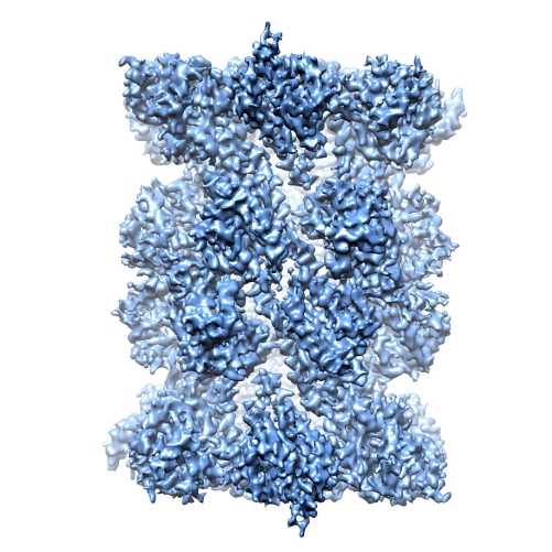 emd_3231.jpg emd_3231.jpg | 191.4 KB | ||
| Archive directory |  http://ftp.pdbj.org/pub/emdb/structures/EMD-3231ftp://ftp.pdbj.org/pub/emdb/structures/EMD-3231 http://ftp.pdbj.org/pub/emdb/structures/EMD-3231ftp://ftp.pdbj.org/pub/emdb/structures/EMD-3231 | HTTPS FTP |
-Related structure data
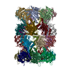
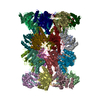
| Related structure data | 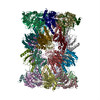 5fmgMC M: atomic model generated by this map C: citing same article ( |
|---|---|
| Similar structure data |


-Links
| EMDB pages | EMDB (EBI/PDBe) / EMDataResource |
|---|---|
| Related items in Molecule of the Month |


-Map
| File | Download / File: emd_3231.map.gz / Format: CCP4 / Size: 62.5 MB / Type: IMAGE STORED AS FLOATING POINT NUMBER (4 BYTES) | ||||||||||||||||||||||||||||||||||||||||||||||||||||||||||||
|---|---|---|---|---|---|---|---|---|---|---|---|---|---|---|---|---|---|---|---|---|---|---|---|---|---|---|---|---|---|---|---|---|---|---|---|---|---|---|---|---|---|---|---|---|---|---|---|---|---|---|---|---|---|---|---|---|---|---|---|---|---|
| Annotation | unsharpened and unfiltered 3D reconstruction of the Plasmodium falciparum 20S proteasome core with a ligand | ||||||||||||||||||||||||||||||||||||||||||||||||||||||||||||
| Voxel size | X=Y=Z: 1.04 Å | ||||||||||||||||||||||||||||||||||||||||||||||||||||||||||||
| Density |
| ||||||||||||||||||||||||||||||||||||||||||||||||||||||||||||
| Symmetry | Space group: 1 | ||||||||||||||||||||||||||||||||||||||||||||||||||||||||||||
| Details | EMDB XML:
CCP4 map header:
| ||||||||||||||||||||||||||||||||||||||||||||||||||||||||||||
-Supplemental data
- Sample components
-Entire : Plasmodium falciparum 20S proteasome core bound to a specific inh...
| Entire | Name: Plasmodium falciparum 20S proteasome core bound to a specific inhibitor |
|---|---|
| Components |
|
-Supramolecule #1000: Plasmodium falciparum 20S proteasome core bound to a specific inh...
| Supramolecule | Name: Plasmodium falciparum 20S proteasome core bound to a specific inhibitor type: sample / ID: 1000 / Number unique components: 2 |
|---|---|
| Molecular weight | Theoretical: 760 KDa |
-Macromolecule #1: proteasome 20S core
| Macromolecule | Name: proteasome 20S core / type: protein_or_peptide / ID: 1 Details: The sample protein components are deposited in the PlasmoDB (the Plasmodium genome resource) Oligomeric state: dimer / Recombinant expression: No |
|---|---|
| Source (natural) | Organism: Plasmodium falciparum (malaria parasite P. falciparum) |
| Molecular weight | Theoretical: 760 KDa |
-Macromolecule #2: Mor-WLW vinyl sulphone (HET: 7F1)
| Macromolecule | Name: Mor-WLW vinyl sulphone (HET: 7F1) / type: ligand / ID: 2 / Recombinant expression: No |
|---|---|
| Source (natural) | Organism: synthetic construct (others) |
-Experimental details
-Structure determination
| Method | cryo EM |
|---|---|
 Processing Processing | single particle reconstruction |
| Aggregation state | particle |
-Sample preparation
| Concentration | 0.1 mg/mL |
|---|---|
| Buffer | pH: 7.5 / Details: 50mM Tris HCl, 5mM MgCl2, 1mM dithiotreitol |
| Grid | Details: 1.2/1.3 Quantifoil, 300 mesh Cu grids, coated with freshly floated thin layer of carbon |
| Vitrification | Cryogen name: ETHANE / Chamber humidity: 95 % / Instrument: FEI VITROBOT MARK III / Method: blot 2.5 seconds before plunging |
- Electron microscopy
| Microscope | FEI TITAN KRIOS |
|---|---|
| Electron beam | Acceleration voltage: 300 kV / Electron source: FIELD EMISSION GUN |
| Electron optics | Illumination mode: FLOOD BEAM / Imaging mode: BRIGHT FIELDBright-field microscopy / Nominal defocus max: 3.2 µm / Nominal defocus min: 1.6 µm / Nominal magnification: 75000 |
| Sample stage | Specimen holder model: FEI TITAN KRIOS AUTOGRID HOLDER |
| Details | Each exposure was recorded as 17 individual frames captured at a rate of 0.056 second/frame, each with an electron dose of 2.8 electrons/square angstrom. Data-set recorded using EPU software. |
| Date | Sep 3, 2014 |
| Image recording | Category: CCD / Film or detector model: FEI FALCON II (4k x 4k) / Number real images: 1816 / Average electron dose: 48 e/Å2 / Details: each image was recorded as 17 frames |
| Experimental equipment |  Model: Titan Krios / Image courtesy: FEI Company |
-Image processing
| CTF correction | Details: full recorded image |
|---|---|
| Final reconstruction | Applied symmetry - Point group: C2 (2 fold cyclic) / Algorithm: OTHER / Resolution.type: BY AUTHOR / Resolution: 3.6 Å / Resolution method: OTHER / Software - Name: Tigris, Spider, Imagic / Number images used: 97720 |
| Details | The particles were selected automatically using Relion, followed by manual inspection for the removal of false positives and addition of false negatives |
-Atomic model buiding 1
| Initial model | PDB ID: |
|---|---|
| Details | The initial models of the Plasmodium falciparum 20S proteasome core subunits were obtained using the PHYRE 2 structure prediction server; model building was done using real space refinement in Coot and Phenix |
| Refinement | Space: REAL / Protocol: FLEXIBLE FIT |
| Output model | PDB-5fmg: |

