 Movie
Movie Controller


+ Open data

- Basic information
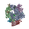
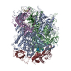
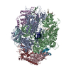
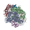
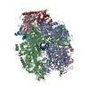
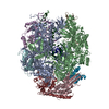
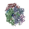
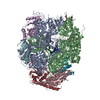
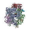
| Entry | Database: EMDB / ID: EMD-3220 | |||||||||
|---|---|---|---|---|---|---|---|---|---|---|
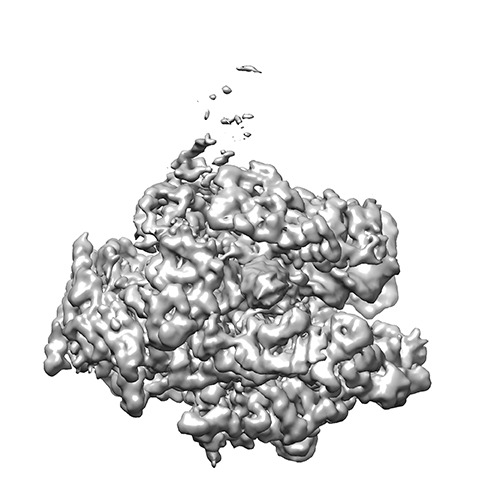
| Title | Structure of transcribing mammalian RNA polymerase II (EC3) | |||||||||
 Map data Map data | Bovine Pol II elongation complex EC3, unsharpened map | |||||||||
 Sample Sample |
| |||||||||
 Keywords Keywords |  transcription / elongation transcription / elongation | |||||||||
| Function / homology | DNA-directed 5'-3' RNA polymerase activity Function and homology information Function and homology information | |||||||||
| Biological species |  Bos taurus (cattle) / Bos taurus (cattle) /  Homo sapiens (human) / unidentified (others) Homo sapiens (human) / unidentified (others) | |||||||||
| Method | single particle reconstruction / cryo EM / Resolution: 3.7 Å | |||||||||
 Authors Authors | Bernecky C / Herzog F / Baumeister W / Plitzko JM / Cramer P | |||||||||
 Citation Citation | Journal: Nature / Year: 2016 Title: Structure of transcribing mammalian RNA polymerase II. Authors: Carrie Bernecky / Franz Herzog / Wolfgang Baumeister / Jürgen M Plitzko / Patrick Cramer /  Abstract: RNA polymerase (Pol) II produces messenger RNA during transcription of protein-coding genes in all eukaryotic cells. The Pol II structure is known at high resolution from X-ray crystallography for ...RNA polymerase (Pol) II produces messenger RNA during transcription of protein-coding genes in all eukaryotic cells. The Pol II structure is known at high resolution from X-ray crystallography for two yeast species. Structural studies of mammalian Pol II, however, remain limited to low-resolution electron microscopy analysis of human Pol II and its complexes with various proteins. Here we report the 3.4 Å resolution cryo-electron microscopy structure of mammalian Pol II in the form of a transcribing complex comprising DNA template and RNA transcript. We use bovine Pol II, which is identical to the human enzyme except for seven amino-acid residues. The obtained atomic model closely resembles its yeast counterpart, but also reveals unknown features. Binding of nucleic acids to the polymerase involves 'induced fit' of the mobile Pol II clamp and active centre region. DNA downstream of the transcription bubble contacts a conserved 'TPSA motif' in the jaw domain of the Pol II subunit RPB5, an interaction that is apparently already established during transcription initiation. Upstream DNA emanates from the active centre cleft at an angle of approximately 105° with respect to downstream DNA. This position of upstream DNA allows for binding of the general transcription elongation factor DSIF (SPT4-SPT5) that we localize over the active centre cleft in a conserved position on the clamp domain of Pol II. Our results define the structure of mammalian Pol II in its functional state, indicate that previous crystallographic analysis of yeast Pol II is relevant for understanding gene transcription in all eukaryotes, and provide a starting point for a mechanistic analysis of human transcription. | |||||||||
| History |
|
- Structure visualization
| Movie |
Movie viewer |
|---|---|
| Structure viewer | EM map: SurfViewMolmilJmol/JSmol |
| Supplemental images |
- Downloads & links
-EMDB archive
| Map data | emd_3220.map.gz | 30 MB | EMDB map data format | |
|---|---|---|---|---|
| Header (meta data) | emd-3220-v30.xmlemd-3220.xml | 10.6 KB 10.6 KB | Display Display | EMDB header |
| Images | emd_3220.tif | 145.4 KB | ||
| Others | EMD-3220_autob.map | 32.4 MB | ||
| Archive directory |  http://ftp.pdbj.org/pub/emdb/structures/EMD-3220ftp://ftp.pdbj.org/pub/emdb/structures/EMD-3220 http://ftp.pdbj.org/pub/emdb/structures/EMD-3220ftp://ftp.pdbj.org/pub/emdb/structures/EMD-3220 | HTTPS FTP |
-Related structure data

-Links
| EMDB pages | EMDB (EBI/PDBe) / EMDataResource |
|---|
-Map
| File | Download / File: emd_3220.map.gz / Format: CCP4 / Size: 31.6 MB / Type: IMAGE STORED AS FLOATING POINT NUMBER (4 BYTES) | ||||||||||||||||||||||||||||||||||||||||||||||||||||||||||||||||||||
|---|---|---|---|---|---|---|---|---|---|---|---|---|---|---|---|---|---|---|---|---|---|---|---|---|---|---|---|---|---|---|---|---|---|---|---|---|---|---|---|---|---|---|---|---|---|---|---|---|---|---|---|---|---|---|---|---|---|---|---|---|---|---|---|---|---|---|---|---|---|
| Annotation | Bovine Pol II elongation complex EC3, unsharpened map | ||||||||||||||||||||||||||||||||||||||||||||||||||||||||||||||||||||
| Voxel size | X=Y=Z: 1.35 Å | ||||||||||||||||||||||||||||||||||||||||||||||||||||||||||||||||||||
| Density |
| ||||||||||||||||||||||||||||||||||||||||||||||||||||||||||||||||||||
| Symmetry | Space group: 1 | ||||||||||||||||||||||||||||||||||||||||||||||||||||||||||||||||||||
| Details | EMDB XML:
CCP4 map header:
| ||||||||||||||||||||||||||||||||||||||||||||||||||||||||||||||||||||
-Supplemental data
-Supplemental map: EMD-3220 autob.map
| File | EMD-3220_autob.map | ||||||||||||
|---|---|---|---|---|---|---|---|---|---|---|---|---|---|
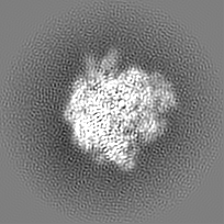
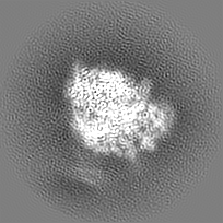
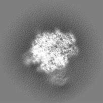
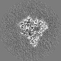
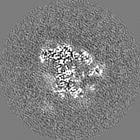
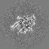
| Projections & Slices |
| ||||||||||||
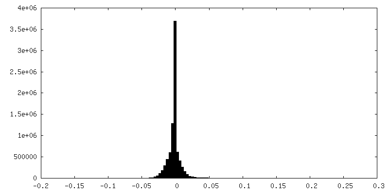
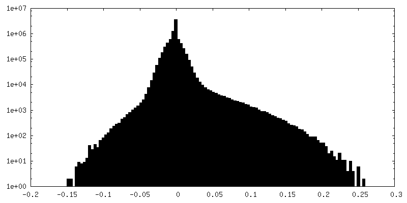
| Density Histograms |
 Z
Z Y
Y X
X

- Sample components
-Entire : bovine Pol II elongation complex
| Entire | Name: bovine Pol II elongation complex |
|---|---|
| Components |
|
-Supramolecule #1000: bovine Pol II elongation complex
| Supramolecule | Name: bovine Pol II elongation complex / type: sample / ID: 1000 Details: Recombinant human Gdown1 was present during sample preparation but density was not observed in this map. Number unique components: 3 |
|---|---|
| Molecular weight | Theoretical: 590 KDa |
-Macromolecule #1: DNA-directed RNA polymerase II
| Macromolecule | Name: DNA-directed RNA polymerase II / type: protein_or_peptide / ID: 1 / Name.synonym: RNA polymerase II / Number of copies: 1 / Oligomeric state: Monomer / Recombinant expression: No |
|---|---|
| Source (natural) | Organism: Bos taurus (cattle) / synonym: Cow / Tissue: Thymus |
| Molecular weight | Theoretical: 520 KDa |
| Sequence | GO: DNA-directed 5'-3' RNA polymerase activity |
-Macromolecule #2: DNA-directed RNA polymerase II subunit GRINL1A
| Macromolecule | Name: DNA-directed RNA polymerase II subunit GRINL1A / type: protein_or_peptide / ID: 2 / Name.synonym: Gdown1 / Number of copies: 1 / Oligomeric state: Monomer / Recombinant expression: Yes |
|---|---|
| Source (natural) | Organism: Homo sapiens (human) / synonym: Human |
| Molecular weight | Theoretical: 40 KDa |
| Recombinant expression | Organism:  Escherichia coli (E. coli) / Recombinant strain: BL21(DE3)RIL / Recombinant plasmid: pOPINB Escherichia coli (E. coli) / Recombinant strain: BL21(DE3)RIL / Recombinant plasmid: pOPINB |
-Macromolecule #3: DNA-RNA synthetic construct
| Macromolecule | Name: DNA-RNA synthetic construct / type: other / ID: 3 / Name.synonym: DNA-RNA elongation scaffold / Classification: DNA/RNA / Structure: OTHER / Synthetic?: Yes |
|---|---|
| Source (natural) | Organism: unidentified (others) |
| Molecular weight | Theoretical: 30 KDa |
-Experimental details
-Structure determination
| Method | cryo EM |
|---|---|
 Processing Processing | single particle reconstruction |
| Aggregation state | particle |
-Sample preparation
| Concentration | 0.3 mg/mL |
|---|---|
| Buffer | pH: 7.25 / Details: 150 mM NaCl, 5 mM HEPES, 0.01 mM ZnCl2, 10 mM DTT |
| Grid | Details: Quantifoil R 3.5/1 holey carbon grids |
| Vitrification | Cryogen name: ETHANE / Chamber humidity: 100 % / Instrument: FEI VITROBOT MARK IV Method: Four microliters of sample was applied to glow-discharged Quantifoil R 3.5/1 holey carbon grids, which were then blotted for 8.5s and plunge-frozen in liquid ethane. |
- Electron microscopy
| Microscope | FEI TITAN KRIOS |
|---|---|
| Electron beam | Acceleration voltage: 300 kV / Electron source: FIELD EMISSION GUN |
| Electron optics | Calibrated magnification: 37037 / Illumination mode: FLOOD BEAM / Imaging mode: BRIGHT FIELDBright-field microscopy / Cs: 2 mm / Nominal defocus max: 3.1 µm / Nominal defocus min: 0.6 µm / Nominal magnification: 37000 |
| Specialist optics | Energy filter - Name: GIF Quantum / Energy filter - Lower energy threshold: 0.0 eV / Energy filter - Upper energy threshold: 20.0 eV |
| Sample stage | Specimen holder model: FEI TITAN KRIOS AUTOGRID HOLDER |
| Date | Dec 1, 2014 |
| Image recording | Category: CCD / Film or detector model: GATAN K2 SUMMIT (4k x 4k) / Number real images: 1172 / Average electron dose: 43 e/Å2 Details: Each movie image was collected over 8 s fractionated into 40 frames (0.2 s each). |
| Experimental equipment |  Model: Titan Krios / Image courtesy: FEI Company |
-Image processing
| CTF correction | Details: Each particle |
|---|---|
| Final reconstruction | Applied symmetry - Point group: C1 (asymmetric) / Algorithm: OTHER / Resolution.type: BY AUTHOR / Resolution: 3.7 Å / Resolution method: OTHER / Software - Name: RELION / Number images used: 184122 |
