 Movie
Movie Controller

[English] 日本語
 Yorodumi
- EMDB-3216: In situ sub-tomogram average of the host-free Chlamydia trachomat... -
+ Open data

- Basic information
| Entry | Database: EMDB / ID: EMD-3216 | |||||||||
|---|---|---|---|---|---|---|---|---|---|---|
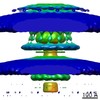
| Title | In situ sub-tomogram average of the host-free Chlamydia trachomatis type III secretion system Type three secretion system Type three secretion system | |||||||||
 Map data Map data | Sub-tomogram average of Chlamydia type III secretion system (host-free)Type three secretion system | |||||||||
 Sample Sample |
| |||||||||
 Keywords Keywords | injectisome / type III secretion / T3SS | |||||||||
| Biological species |   Chlamydia trachomatis (bacteria) Chlamydia trachomatis (bacteria) | |||||||||
| Method | subtomogram averaging / cryo EM / Resolution: 33.0 Å | |||||||||
 Authors Authors | Nans A / Kudryashev M / Saibil HR / Hayward RD | |||||||||
 Citation Citation | Journal: Nat Commun / Year: 2015 Title: Structure of a bacterial type III secretion system in contact with a host membrane in situ. Authors: Andrea Nans / Mikhail Kudryashev / Helen R Saibil / Richard D Hayward /   Abstract: Many bacterial pathogens of animals and plants use a conserved type III secretion system (T3SS) to inject virulence effector proteins directly into eukaryotic cells to subvert host functions. Contact ...Many bacterial pathogens of animals and plants use a conserved type III secretion system (T3SS) to inject virulence effector proteins directly into eukaryotic cells to subvert host functions. Contact with host membranes is critical for T3SS activation, yet little is known about T3SS architecture in this state or the conformational changes that drive effector translocation. Here we use cryo-electron tomography and sub-tomogram averaging to derive the intact structure of the primordial Chlamydia trachomatis T3SS in the presence and absence of host membrane contact. Comparison of the averaged structures demonstrates a marked compaction of the basal body (4 nm) occurs when the needle tip contacts the host cell membrane. This compaction is coupled to a stabilization of the cytosolic sorting platform-ATPase. Our findings reveal the first structure of a bacterial T3SS from a major human pathogen engaged with a eukaryotic host, and reveal striking 'pump-action' conformational changes that underpin effector injection. | |||||||||
| History |
|
- Structure visualization
| Movie |
 Movie viewer Movie viewer |
|---|---|
| Structure viewer | EM map: SurfViewMolmilJmol/JSmol |
| Supplemental images |

- Downloads & links
-EMDB archive
| Map data | emd_3216.map.gz | 41 MB | EMDB map data format | |
|---|---|---|---|---|
| Header (meta data) | emd-3216-v30.xmlemd-3216.xml | 8.2 KB 8.2 KB | Display Display | EMDB header |
| Images | EMD-3216_snapshot36_500.tif | 978.8 KB | ||
| Others | Subtomo_EMD-3216.map.gz | 12 MB | ||
| Archive directory |  http://ftp.pdbj.org/pub/emdb/structures/EMD-3216ftp://ftp.pdbj.org/pub/emdb/structures/EMD-3216 http://ftp.pdbj.org/pub/emdb/structures/EMD-3216ftp://ftp.pdbj.org/pub/emdb/structures/EMD-3216 | HTTPS FTP |
-Related structure data
| Related structure data |  3217C C: citing same article ( |
|---|---|
| Similar structure data | |
| EM raw data | EMPIAR-10047 (Title: Cryo-electron tomogram of host-free Chlamydia trachomatis with type III secretion system Data size: 11.6 Data #1: Aligned multi-frame micrographs that comprise a tilt series of Chlamydia trachomatis type III secretion systems [class averages]) |


-Links
| EMDB pages | EMDB (EBI/PDBe) / EMDataResource |
|---|
-Map
| File | Download / File: emd_3216.map.gz / Format: CCP4 / Size: 62.5 MB / Type: IMAGE STORED AS FLOATING POINT NUMBER (4 BYTES) | ||||||||||||||||||||||||||||||||||||||||||||||||||||||||||||||||||||
|---|---|---|---|---|---|---|---|---|---|---|---|---|---|---|---|---|---|---|---|---|---|---|---|---|---|---|---|---|---|---|---|---|---|---|---|---|---|---|---|---|---|---|---|---|---|---|---|---|---|---|---|---|---|---|---|---|---|---|---|---|---|---|---|---|---|---|---|---|---|
| Annotation | Sub-tomogram average of Chlamydia type III secretion system (host-free) | ||||||||||||||||||||||||||||||||||||||||||||||||||||||||||||||||||||
| Voxel size | X=Y=Z: 5.4 Å | ||||||||||||||||||||||||||||||||||||||||||||||||||||||||||||||||||||
| Density |
| ||||||||||||||||||||||||||||||||||||||||||||||||||||||||||||||||||||
| Symmetry | Space group: 1 | ||||||||||||||||||||||||||||||||||||||||||||||||||||||||||||||||||||
| Details | EMDB XML:
CCP4 map header:
| ||||||||||||||||||||||||||||||||||||||||||||||||||||||||||||||||||||
-Supplemental data
-Supplemental map: Subtomo EMD-3216.map
| File | Subtomo_EMD-3216.map | ||||||||||||
|---|---|---|---|---|---|---|---|---|---|---|---|---|---|
| Projections & Slices |
| ||||||||||||
| Density Histograms |
 Z
Z Y
Y X
X


- Sample components
-Entire : Chlamydia trachomatis type III secretion system (host-free)
| Entire | Name: Chlamydia trachomatis type III secretion system (host-free)Type three secretion system |
|---|---|
| Components |
|
-Supramolecule #1000: Chlamydia trachomatis type III secretion system (host-free)
| Supramolecule | Name: Chlamydia trachomatis type III secretion system (host-free) type: sample / ID: 1000 / Number unique components: 1 |
|---|
-Supramolecule #1: Chlamydia trachomatis type III secretion system (host-free)
| Supramolecule | Name: Chlamydia trachomatis type III secretion system (host-free) type: organelle_or_cellular_component / ID: 1 / Recombinant expression: No |
|---|---|
| Source (natural) | Organism: Chlamydia trachomatis (bacteria) |
-Experimental details
-Structure determination
| Method | cryo EM |
|---|---|
 Processing Processing | subtomogram averaging |
| Aggregation state | cell |
-Sample preparation
| Grid | Details: 200 mesh gold quantifoil grid 3.5/1 |
|---|---|
| Vitrification | Cryogen name: ETHANE / Chamber humidity: 100 % / Instrument: FEI VITROBOT MARK IV / Method: Blot for 2.5 seconds |
- Electron microscopy
| Microscope | FEI POLARA 300 |
|---|---|
| Electron beam | Acceleration voltage: 300 kV / Electron source: FIELD EMISSION GUN |
| Electron optics | Illumination mode: FLOOD BEAM / Imaging mode: BRIGHT FIELDBright-field microscopy / Cs: 2.3 mm / Nominal defocus max: -10.0 µm / Nominal magnification: 41000 |
| Specialist optics | Energy filter - Name: Gatan Quantum / Energy filter - Lower energy threshold: 0.0 eV / Energy filter - Upper energy threshold: 20.0 eV |
| Sample stage | Specimen holder model: SIDE ENTRY, EUCENTRIC / Tilt series - Axis1 - Min angle: -45 ° / Tilt series - Axis1 - Max angle: 60 ° |
| Date | Sep 1, 2014 |
| Image recording | Category: CCD / Film or detector model: GATAN K2 SUMMIT (4k x 4k) / Average electron dose: 55 e/Å2 |
| Experimental equipment |  Model: Tecnai Polara / Image courtesy: FEI Company |
-Image processing
| CTF correction | Details: Each micrograph was phase-flipped in IMOD according to the measured defocus. |
|---|---|
| Final reconstruction | Applied symmetry - Point group: C12 (12 fold cyclic) / Resolution.type: BY AUTHOR / Resolution: 33.0 Å / Resolution method: OTHER / Software - Name: IMOD, Dynamo / Number subtomograms used: 515 |
| Details | Sub-tomograms selected in IMOD and cropped with Dynamo. Alignment and averaging was done in Dynamo. |
