 Movie
Movie Controller

[English] 日本語
 Yorodumi
- EMDB-3100: Cryo-electron tomography of a Golgi intracisternal protein array -
+ Open data

- Basic information
| Entry | Database: EMDB / ID: EMD-3100 | |||||||||
|---|---|---|---|---|---|---|---|---|---|---|
| Title | Cryo-electron tomography of a Golgi intracisternal protein array | |||||||||
 Map data Map data | Symmetrized average of a trans-Golgi intracisternal protein array | |||||||||
 Sample Sample |
| |||||||||
 Keywords Keywords | Golgi /  transmembrane protein / cisterna / focused ion beam / tomography transmembrane protein / cisterna / focused ion beam / tomography | |||||||||
| Biological species |   Chlamydomonas reinhardtii (plant) Chlamydomonas reinhardtii (plant) | |||||||||
| Method | subtomogram averaging / cryo EM / Resolution: 27.0 Å | |||||||||
 Authors Authors | Engel BD / Schaffer M / Albert S / Asano S / Plitzko JM / Baumeister W | |||||||||
 Citation Citation | Journal: Proc Natl Acad Sci U S A / Year: 2015 Title: In situ structural analysis of Golgi intracisternal protein arrays. Authors: Benjamin D Engel / Miroslava Schaffer / Sahradha Albert / Shoh Asano / Jürgen M Plitzko / Wolfgang Baumeister /  Abstract: We acquired molecular-resolution structures of the Golgi within its native cellular environment. Vitreous Chlamydomonas cells were thinned by cryo-focused ion beam milling and then visualized by cryo- ...We acquired molecular-resolution structures of the Golgi within its native cellular environment. Vitreous Chlamydomonas cells were thinned by cryo-focused ion beam milling and then visualized by cryo-electron tomography. These tomograms revealed structures within the Golgi cisternae that have not been seen before. Narrow trans-Golgi lumina were spanned by asymmetric membrane-associated protein arrays that had ∼6-nm lateral periodicity. Subtomogram averaging showed that the arrays may determine the narrow central spacing of the trans-Golgi cisternae through zipper-like interactions, thereby forcing cargo to the trans-Golgi periphery. Additionally, we observed dense granular aggregates within cisternae and intracisternal filament bundles associated with trans-Golgi buds. These native in situ structures provide new molecular insights into Golgi architecture and function. | |||||||||
| History |
|
- Structure visualization
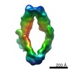
| Movie |
 Movie viewer Movie viewer |
|---|---|
| Structure viewer | EM map: SurfViewMolmilJmol/JSmol |
| Supplemental images |


- Downloads & links
-EMDB archive
| Map data | emd_3100.map.gz | 1.8 MB | EMDB map data format | |
|---|---|---|---|---|
| Header (meta data) | emd-3100-v30.xmlemd-3100.xml | 8.3 KB 8.3 KB | Display Display | EMDB header |
| Images | 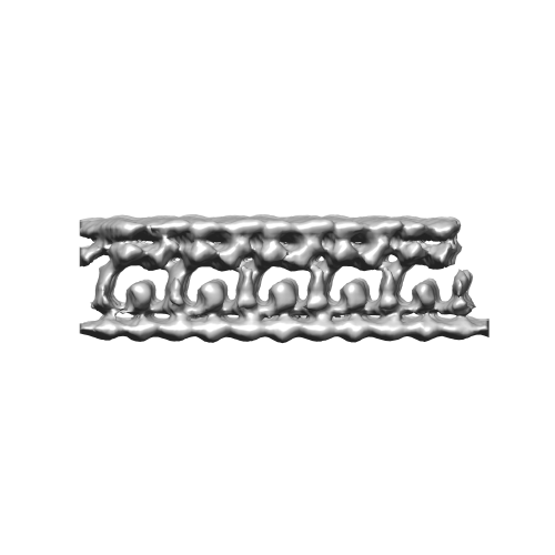 EMD-3100.png EMD-3100.png emd_3100.png emd_3100.png | 49.6 KB 49.6 KB | ||
| Archive directory |  http://ftp.pdbj.org/pub/emdb/structures/EMD-3100ftp://ftp.pdbj.org/pub/emdb/structures/EMD-3100 http://ftp.pdbj.org/pub/emdb/structures/EMD-3100ftp://ftp.pdbj.org/pub/emdb/structures/EMD-3100 | HTTPS FTP |
-Related structure data
| Similar structure data |
|---|
-Links
| EMDB pages | EMDB (EBI/PDBe) / EMDataResource |
|---|
-Map
| File | Download / File: emd_3100.map.gz / Format: CCP4 / Size: 3.3 MB / Type: IMAGE STORED AS FLOATING POINT NUMBER (4 BYTES) | ||||||||||||||||||||||||||||||||||||||||||||||||||||||||||||||||||||
|---|---|---|---|---|---|---|---|---|---|---|---|---|---|---|---|---|---|---|---|---|---|---|---|---|---|---|---|---|---|---|---|---|---|---|---|---|---|---|---|---|---|---|---|---|---|---|---|---|---|---|---|---|---|---|---|---|---|---|---|---|---|---|---|---|---|---|---|---|---|
| Annotation | Symmetrized average of a trans-Golgi intracisternal protein array | ||||||||||||||||||||||||||||||||||||||||||||||||||||||||||||||||||||
| Voxel size | X=Y=Z: 6.84 Å | ||||||||||||||||||||||||||||||||||||||||||||||||||||||||||||||||||||
| Density |
| ||||||||||||||||||||||||||||||||||||||||||||||||||||||||||||||||||||
| Symmetry | Space group: 1 | ||||||||||||||||||||||||||||||||||||||||||||||||||||||||||||||||||||
| Details | EMDB XML:
CCP4 map header:
| ||||||||||||||||||||||||||||||||||||||||||||||||||||||||||||||||||||
-Supplemental data
- Sample components
-Entire : Unknown protein complex linking the cisternal membranes of the tr...
| Entire | Name: Unknown protein complex linking the cisternal membranes of the trans-Golgi |
|---|---|
| Components |
|
-Supramolecule #1000: Unknown protein complex linking the cisternal membranes of the tr...
| Supramolecule | Name: Unknown protein complex linking the cisternal membranes of the trans-Golgi type: sample / ID: 1000 / Number unique components: 1 |
|---|
-Supramolecule #1: trans-Golgi intracisternal protein array
| Supramolecule | Name: trans-Golgi intracisternal protein array / type: organelle_or_cellular_component / ID: 1 / Recombinant expression: No |
|---|---|
| Source (natural) | Organism: Chlamydomonas reinhardtii (plant) / Organelle: Golgi / Location in cell: trans-Golgi cisternae |
-Experimental details
-Structure determination
| Method | cryo EM |
|---|---|
 Processing Processing | subtomogram averaging |
| Aggregation state | cell |
-Sample preparation
| Vitrification | Cryogen name: ETHANE-PROPANE MIXTURE / Chamber humidity: 95 % / Instrument: FEI VITROBOT MARK IV Details: Focused ion-beam milling was used to thin the sample |
|---|
- Electron microscopy
| Microscope | FEI TITAN KRIOS |
|---|---|
| Electron beam | Acceleration voltage: 300 kV / Electron source: FIELD EMISSION GUN |
| Electron optics | Calibrated magnification: 14600 / Illumination mode: FLOOD BEAM / Imaging mode: BRIGHT FIELDBright-field microscopy / Cs: 2 mm / Nominal defocus max: -0.006 µm / Nominal defocus min: -0.005 µm / Nominal magnification: 42000 |
| Specialist optics | Energy filter - Name: Gatan |
| Sample stage | Specimen holder model: FEI TITAN KRIOS AUTOGRID HOLDER / Tilt series - Axis1 - Min angle: -60 ° / Tilt series - Axis1 - Max angle: 60 ° |
| Date | Feb 20, 2015 |
| Image recording | Category: CCD / Film or detector model: GATAN K2 (4k x 4k) / Number real images: 60 / Average electron dose: 60 e/Å2 |
| Experimental equipment |  Model: Titan Krios / Image courtesy: FEI Company |
-Image processing
| CTF correction | Details: each tilt |
|---|---|
| Final reconstruction | Applied symmetry - Point group: C1 (asymmetric) / Algorithm: OTHER / Resolution.type: BY AUTHOR / Resolution: 27.0 Å / Resolution method: OTHER / Software - Name: IMOD, TOM, pyTOM / Number subtomograms used: 244 |
| Details | translational symmetry was applied |
