 Movie
Movie Controller

[English] 日本語
 Yorodumi
- EMDB-3079: Single-particle cryo-EM of co-translational folded adr1 domain in... -
+ Open data

- Basic information
| Entry | Database: EMDB / ID: EMD-3079 | |||||||||
|---|---|---|---|---|---|---|---|---|---|---|
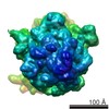
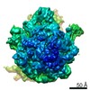
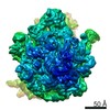
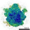
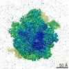
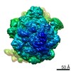
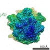
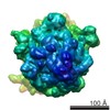
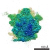
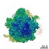
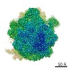
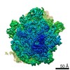
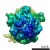
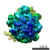
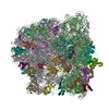
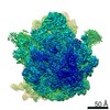
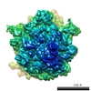
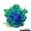
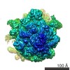
| Title | Single-particle cryo-EM of co-translational folded adr1 domain inside the E. coli ribosome exit tunnel. | |||||||||
 Map data Map data | Adr1 domain inside the ribosome exit tunnel | |||||||||
 Sample Sample |
| |||||||||
 Keywords Keywords |  protein folding / translation / ribosome / zinc finger / SecM / translational arrest peptide / cryo-EM / single-molecule studies protein folding / translation / ribosome / zinc finger / SecM / translational arrest peptide / cryo-EM / single-molecule studies | |||||||||
| Function / homology |  Function and homology information Function and homology informationpositive regulation of transcription from RNA polymerase II promoter by oleic acid / peroxisome organization / TFIID-class transcription factor complex binding / TFIIB-class transcription factor binding / chromatin organization / RNA polymerase II-specific DNA-binding transcription factor binding / sequence-specific DNA binding / nucleic acid binding / transcription coactivator activity / molecular adaptor activity ...positive regulation of transcription from RNA polymerase II promoter by oleic acid / peroxisome organization / TFIID-class transcription factor complex binding / TFIIB-class transcription factor binding / chromatin organization / RNA polymerase II-specific DNA-binding transcription factor binding / sequence-specific DNA binding / nucleic acid binding / transcription coactivator activity / molecular adaptor activity / DNA-binding transcription factor activity, RNA polymerase II-specific / RNA polymerase II cis-regulatory region sequence-specific DNA binding / chromatin / regulation of transcription by RNA polymerase II / positive regulation of transcription by RNA polymerase II / metal ion binding / nucleus / cytosol / cytoplasmSimilarity search - Function | |||||||||
| Biological species |  Escherichia coli (E. coli) / Escherichia coli (E. coli) /  Saccharomyces cerevisiae (brewer's yeast) Saccharomyces cerevisiae (brewer's yeast) | |||||||||
| Method | single particle reconstruction / cryo EM / Resolution: 4.8 Å | |||||||||
 Authors Authors | Nilsson OB / Hedman R / Marino J / Wickles S / Bischoff L / Johansson M / Muller-Lucks A / Trovato F / Puglisi JD / O'Brien E ...Nilsson OB / Hedman R / Marino J / Wickles S / Bischoff L / Johansson M / Muller-Lucks A / Trovato F / Puglisi JD / O'Brien E / Beckmann R / von Heijne G | |||||||||
 Citation Citation | Journal: Cell Rep / Year: 2015 Title: Cotranslational Protein Folding inside the Ribosome Exit Tunnel. Authors: Ola B Nilsson / Rickard Hedman / Jacopo Marino / Stephan Wickles / Lukas Bischoff / Magnus Johansson / Annika Müller-Lucks / Fabio Trovato / Joseph D Puglisi / Edward P O'Brien / Roland ...Authors: Ola B Nilsson / Rickard Hedman / Jacopo Marino / Stephan Wickles / Lukas Bischoff / Magnus Johansson / Annika Müller-Lucks / Fabio Trovato / Joseph D Puglisi / Edward P O'Brien / Roland Beckmann / Gunnar von Heijne /   Abstract: At what point during translation do proteins fold? It is well established that proteins can fold cotranslationally outside the ribosome exit tunnel, whereas studies of folding inside the exit tunnel ...At what point during translation do proteins fold? It is well established that proteins can fold cotranslationally outside the ribosome exit tunnel, whereas studies of folding inside the exit tunnel have so far detected only the formation of helical secondary structure and collapsed or partially structured folding intermediates. Here, using a combination of cotranslational nascent chain force measurements, inter-subunit fluorescence resonance energy transfer studies on single translating ribosomes, molecular dynamics simulations, and cryoelectron microscopy, we show that a small zinc-finger domain protein can fold deep inside the vestibule of the ribosome exit tunnel. Thus, for small protein domains, the ribosome itself can provide the kind of sheltered folding environment that chaperones provide for larger proteins. | |||||||||
| History |
|
- Structure visualization
| Movie |
Movie viewer |
|---|---|
| Structure viewer | EM map: SurfViewMolmilJmol/JSmol |
| Supplemental images |

- Downloads & links
-EMDB archive
| Map data | emd_3079.map.gz | 177.6 MB | EMDB map data format | |
|---|---|---|---|---|
| Header (meta data) | emd-3079-v30.xmlemd-3079.xml | 9.7 KB 9.7 KB | Display Display | EMDB header |
| Images | EMD-3079.tif | 136.9 KB | ||
| Archive directory |  http://ftp.pdbj.org/pub/emdb/structures/EMD-3079ftp://ftp.pdbj.org/pub/emdb/structures/EMD-3079 http://ftp.pdbj.org/pub/emdb/structures/EMD-3079ftp://ftp.pdbj.org/pub/emdb/structures/EMD-3079 | HTTPS FTP |
-Related structure data
| Related structure data |  5a7uMC M: atomic model generated by this map C: citing same article ( |
|---|---|
| Similar structure data |
-Links
| EMDB pages | EMDB (EBI/PDBe) / EMDataResource |
|---|---|
| Related items in Molecule of the Month |


-Map
| File | Download / File: emd_3079.map.gz / Format: CCP4 / Size: 185.7 MB / Type: IMAGE STORED AS FLOATING POINT NUMBER (4 BYTES) | ||||||||||||||||||||||||||||||||||||||||||||||||||||||||||||||||||||
|---|---|---|---|---|---|---|---|---|---|---|---|---|---|---|---|---|---|---|---|---|---|---|---|---|---|---|---|---|---|---|---|---|---|---|---|---|---|---|---|---|---|---|---|---|---|---|---|---|---|---|---|---|---|---|---|---|---|---|---|---|---|---|---|---|---|---|---|---|---|
| Annotation | Adr1 domain inside the ribosome exit tunnel | ||||||||||||||||||||||||||||||||||||||||||||||||||||||||||||||||||||
| Voxel size | X=Y=Z: 1.37 Å | ||||||||||||||||||||||||||||||||||||||||||||||||||||||||||||||||||||
| Density |
| ||||||||||||||||||||||||||||||||||||||||||||||||||||||||||||||||||||
| Symmetry | Space group: 1 | ||||||||||||||||||||||||||||||||||||||||||||||||||||||||||||||||||||
| Details | EMDB XML:
CCP4 map header:
| ||||||||||||||||||||||||||||||||||||||||||||||||||||||||||||||||||||
-Supplemental data
- Sample components
-Entire : adr1 domain cotranslationally folded inside the E. coli ribosome ...
| Entire | Name: adr1 domain cotranslationally folded inside the E. coli ribosome exit tunnel |
|---|---|
| Components |
|
-Supramolecule #1000: adr1 domain cotranslationally folded inside the E. coli ribosome ...
| Supramolecule | Name: adr1 domain cotranslationally folded inside the E. coli ribosome exit tunnel type: sample / ID: 1000 / Oligomeric state: One monomer of adr1 domain with ribosome / Number unique components: 2 |
|---|
-Supramolecule #1: 70s ribosome
| Supramolecule | Name: 70s ribosome / type: complex / ID: 1 / Recombinant expression: No / Ribosome-details: ribosome-prokaryote: ALL |
|---|---|
| Source (natural) | Organism: Escherichia coli (E. coli) |
-Macromolecule #1: adr1 domain
| Macromolecule | Name: adr1 domain / type: protein_or_peptide / ID: 1 / Number of copies: 1 / Oligomeric state: Monomer / Recombinant expression: Yes |
|---|---|
| Source (natural) | Organism: Saccharomyces cerevisiae (brewer's yeast) / synonym: Baker's Yeast |
| Molecular weight | Theoretical: 151 KDa |
| Recombinant expression | Organism: Escherichia coli (E. coli) |
| Sequence | UniProtKB: Regulatory protein ADR1 |
-Experimental details
-Structure determination
| Method | cryo EM |
|---|---|
 Processing Processing | single particle reconstruction |
| Aggregation state | particle |
-Sample preparation
| Buffer | pH: 7.2 Details: 20 mM Hepes pH 7.2 , 50 mM KOAc, 5 mM Mg[OAc]2, 0.03% DDM, 50 microM ZnCl2, 125 mM sucrose. |
|---|---|
| Vitrification | Cryogen name: ETHANE / Instrument: FEI VITROBOT MARK IV |
- Electron microscopy
| Microscope | FEI TITAN KRIOS |
|---|---|
| Electron beam | Acceleration voltage: 300 kV / Electron source: FIELD EMISSION GUN |
| Electron optics | Illumination mode: SPOT SCAN / Imaging mode: BRIGHT FIELDBright-field microscopy / Nominal defocus max: -3.2 µm / Nominal defocus min: -1.0 µm |
| Sample stage | Specimen holder model: FEI TITAN KRIOS AUTOGRID HOLDER |
| Date | Apr 13, 2015 |
| Image recording | Category: CCD / Film or detector model: FEI FALCON II (4k x 4k) / Number real images: 3308 / Average electron dose: 5 e/Å2 |
| Experimental equipment |  Model: Titan Krios / Image courtesy: FEI Company |
-Image processing
| CTF correction | Details: Micrograph |
|---|---|
| Final reconstruction | Applied symmetry - Point group: C1 (asymmetric) / Resolution.type: BY AUTHOR / Resolution: 4.8 Å / Resolution method: OTHER / Software - Name: Spider / Number images used: 151900 |

