 Movie
Movie Controller

[English] 日本語
 Yorodumi
- EMDB-3027: Electron cryo-microscopy of porcine Factor VIII bound to lipid na... -
+ Open data

- Basic information
| Entry | Database: EMDB / ID: EMD-3027 | |||||||||
|---|---|---|---|---|---|---|---|---|---|---|
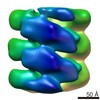
| Title | Electron cryo-microscopy of porcine Factor VIII bound to lipid nanotubes and helical reconstruction | |||||||||
 Map data Map data | Helical reconstruction of porcine Factor VIII bound to LNT as described in the submitted references. | |||||||||
 Sample Sample |
| |||||||||
 Keywords Keywords | Blood coagulation factor VIII / Lipid nanotubes /  Cryo-electron microscopy / Membrane-bound organization / Helical reconstruction Cryo-electron microscopy / Membrane-bound organization / Helical reconstruction | |||||||||
| Biological species |  Sus scrofa (pig) / unidentified (others) Sus scrofa (pig) / unidentified (others) | |||||||||
| Method | helical reconstruction / cryo EM / negative staining / Resolution: 15.5 Å | |||||||||
 Authors Authors | Dalm D / Galaz-Montoya JG / Miller JL / Grushin K / Villalobos A / Koyfman AY / Schmid MF / Stoilova-McPhie S | |||||||||
 Citation Citation | Journal: J Vis Exp / Year: 2014 Title: Helical organization of blood coagulation factor VIII on lipid nanotubes. Authors: Jaimy Miller / Daniela Dalm / Alexey Y Koyfman / Kirill Grushin / Svetla Stoilova-McPhie /  Abstract: Cryo-electron microscopy (Cryo-EM)(1) is a powerful approach to investigate the functional structure of proteins and complexes in a hydrated state and membrane environment(2). Coagulation Factor VIII ...Cryo-electron microscopy (Cryo-EM)(1) is a powerful approach to investigate the functional structure of proteins and complexes in a hydrated state and membrane environment(2). Coagulation Factor VIII (FVIII)(3) is a multi-domain blood plasma glycoprotein. Defect or deficiency of FVIII is the cause for Hemophilia type A - a severe bleeding disorder. Upon proteolytic activation, FVIII binds to the serine protease Factor IXa on the negatively charged platelet membrane, which is critical for normal blood clotting(4). Despite the pivotal role FVIII plays in coagulation, structural information for its membrane-bound state is incomplete(5). Recombinant FVIII concentrate is the most effective drug against Hemophilia type A and commercially available FVIII can be expressed as human or porcine, both forming functional complexes with human Factor IXa(6,7). In this study we present a combination of Cryo-electron microscopy (Cryo-EM), lipid nanotechnology and structure analysis applied to resolve the membrane-bound structure of two highly homologous FVIII forms: human and porcine. The methodology developed in our laboratory to helically organize the two functional recombinant FVIII forms on negatively charged lipid nanotubes (LNT) is described. The representative results demonstrate that our approach is sufficiently sensitive to define the differences in the helical organization between the two highly homologous in sequence (86% sequence identity) proteins. Detailed protocols for the helical organization, Cryo-EM and electron tomography (ET) data acquisition are given. The two-dimensional (2D) and three-dimensional (3D) structure analysis applied to obtain the 3D reconstructions of human and porcine FVIII-LNT is discussed. The presented human and porcine FVIII-LNT structures show the potential of the proposed methodology to calculate the functional, membrane-bound organization of blood coagulation Factor VIII at high resolution. | |||||||||
| History |
|
- Structure visualization
| Movie |
 Movie viewer Movie viewer |
|---|---|
| Structure viewer | EM map: SurfViewMolmilJmol/JSmol |
| Supplemental images |

- Downloads & links
-EMDB archive
| Map data | emd_3027.map.gz | 10.7 MB | EMDB map data format | |
|---|---|---|---|---|
| Header (meta data) | emd-3027-v30.xmlemd-3027.xml | 12.4 KB 12.4 KB | Display Display | EMDB header |
| Images | 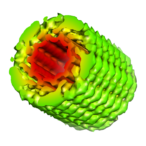 image3027.png image3027.png | 188.6 KB | ||
| Archive directory |  http://ftp.pdbj.org/pub/emdb/structures/EMD-3027ftp://ftp.pdbj.org/pub/emdb/structures/EMD-3027 http://ftp.pdbj.org/pub/emdb/structures/EMD-3027ftp://ftp.pdbj.org/pub/emdb/structures/EMD-3027 | HTTPS FTP |
-Related structure data


-Links
| EMDB pages | EMDB (EBI/PDBe) / EMDataResource |
|---|
-Map
| File | Download / File: emd_3027.map.gz / Format: CCP4 / Size: 41.4 MB / Type: IMAGE STORED AS FLOATING POINT NUMBER (4 BYTES) | ||||||||||||||||||||||||||||||||||||||||||||||||||||||||||||||||||||
|---|---|---|---|---|---|---|---|---|---|---|---|---|---|---|---|---|---|---|---|---|---|---|---|---|---|---|---|---|---|---|---|---|---|---|---|---|---|---|---|---|---|---|---|---|---|---|---|---|---|---|---|---|---|---|---|---|---|---|---|---|---|---|---|---|---|---|---|---|---|
| Annotation | Helical reconstruction of porcine Factor VIII bound to LNT as described in the submitted references. | ||||||||||||||||||||||||||||||||||||||||||||||||||||||||||||||||||||
| Voxel size | X=Y=Z: 2.9 Å | ||||||||||||||||||||||||||||||||||||||||||||||||||||||||||||||||||||
| Density |
| ||||||||||||||||||||||||||||||||||||||||||||||||||||||||||||||||||||
| Symmetry | Space group: 1 | ||||||||||||||||||||||||||||||||||||||||||||||||||||||||||||||||||||
| Details | EMDB XML:
CCP4 map header:
| ||||||||||||||||||||||||||||||||||||||||||||||||||||||||||||||||||||
-Supplemental data
- Sample components
-Entire : Helically organized porcine blood coagulation Factor VIII bound t...
| Entire | Name: Helically organized porcine blood coagulation Factor VIII bound to a lipid nanotube |
|---|---|
| Components |
|
-Supramolecule #1000: Helically organized porcine blood coagulation Factor VIII bound t...
| Supramolecule | Name: Helically organized porcine blood coagulation Factor VIII bound to a lipid nanotube type: sample / ID: 1000 / Oligomeric state: dimer / Number unique components: 2 |
|---|---|
| Molecular weight | Experimental: 170 KDa / Theoretical: 170 KDa / Method: SDS gel and amino acid sequence |
-Macromolecule #1: Blood coagulation Factor VIII
| Macromolecule | Name: Blood coagulation Factor VIII / type: protein_or_peptide / ID: 1 / Name.synonym: Hemophilia A factor Details: Recombinant porcine Factor VIII lacking the B domain was bound to lipid nanotubes and helically organized for structure analysis by cryo-EM. Number of copies: 2 / Oligomeric state: dimer / Recombinant expression: Yes |
|---|---|
| Source (natural) | Organism: Sus scrofa (pig) / synonym: PIG / Cell: blood |
| Molecular weight | Experimental: 170 KDa / Theoretical: 170 KDa |
| Recombinant expression | Organism:  Cricetulus griseus (Chinese hamster) / Recombinant cell: BHK cells Cricetulus griseus (Chinese hamster) / Recombinant cell: BHK cells |
-Macromolecule #2: lipid nanotubes
| Macromolecule | Name: lipid nanotubes / type: ligand / ID: 2 / Recombinant expression: No / Database: NCBI |
|---|---|
| Source (natural) | Organism: unidentified (others) |
-Experimental details
-Structure determination
| Method | negative staining, cryo EM |
|---|---|
 Processing Processing | helical reconstruction |
| Aggregation state | helical array |
-Sample preparation
| Concentration | 1.0 mg/mL |
|---|---|
| Buffer | pH: 7.4 / Details: 20 mM HEPES, 150 mM NaCL, 5 mM CaCl2 |
| Staining | Type: NEGATIVE Details: samples are adsorbed on quantifoil grids and flash frozen in liquid ethane |
| Grid | Details: carbon coated quantifoil grids Q2x2 were glow discharged for 100 second |
| Vitrification | Cryogen name: ETHANE / Chamber temperature: 85 K / Instrument: FEI VITROBOT MARK IV / Method: blot for 3.5 seconds, force 1 before plunging |
| Details | The protein and lipid are mixed in 1:1 ration and incubated for 15 minutes |
- Electron microscopy
| Microscope | JEOL 2100 |
|---|---|
| Electron beam | Acceleration voltage: 200 kV / Electron source: LAB6 |
| Electron optics | Calibrated magnification: 52000 / Illumination mode: FLOOD BEAM / Imaging mode: BRIGHT FIELDBright-field microscopy / Cs: 2.0 mm / Nominal defocus max: 0.004 µm / Nominal defocus min: 0.001 µm / Nominal magnification: 40000 |
| Sample stage | Specimen holder model: GATAN LIQUID NITROGEN |
| Temperature | Average: 99 K |
| Date | Sep 11, 2013 |
| Image recording | Category: CCD / Film or detector model: GATAN ULTRASCAN 4000 (4k x 4k) / Number real images: 300 / Average electron dose: 16 e/Å2 |
-Image processing
| CTF correction | Details: each particle set |
|---|---|
| Final reconstruction | Applied symmetry - Helical parameters - Δz: 36 Å Applied symmetry - Helical parameters - Δ&Phi: 35.5 ° Applied symmetry - Helical parameters - Axial symmetry: C5 (5 fold cyclic )Resolution.type: BY AUTHOR / Resolution: 15.5 Å / Resolution method: OTHER |
| Details | Described in supplemental materials and references |
-Atomic model buiding 1
| Initial model | PDB ID: Chain - #0 - Chain ID: A / Chain - #1 - Chain ID: B |
|---|---|
| Software | Name: UCSF Chimera |
| Refinement | Space: REAL / Protocol: RIGID BODY FIT |

