[English] 日本語
Yorodumi
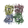
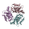
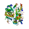
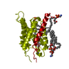
- PDB-2zz9: Structure of aquaporin-4 S180D mutant at 2.8 A resolution by elec... -

+
Open data


ID or keywords:

Loading...

-
Basic information

Entry
Database: PDB / ID: 2zz9
Title
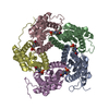
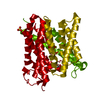
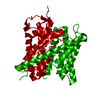
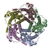
Structure of aquaporin-4 S180D mutant at 2.8 A resolution by electron crystallography
ComponentsAquaporin-4
KeywordsTRANSPORT PROTEIN / WATER TRANSPORT / WATER CHANNEL / AQUAPORIN / TWO-DIMENSIONAL CRYSTAL / MEMBRANE PROTEIN / BACULOVIRUS EXPRESSION SYSTEM / Glycoprotein / Membrane / Phosphoprotein / Transmembrane / Transport
Function / homology
Function and homology information


Passive transport by Aquaporins / cerebrospinal fluid secretion / renal water absorption / regulation of vascular endothelial growth factor production / cerebrospinal fluid circulation / astrocyte end-foot / water channel activity / intracellular water homeostasis / water transport / negative regulation of cell adhesion molecule production ...Passive transport by Aquaporins / cerebrospinal fluid secretion / renal water absorption / regulation of vascular endothelial growth factor production / cerebrospinal fluid circulation / astrocyte end-foot / water channel activity / intracellular water homeostasis / water transport / negative regulation of cell adhesion molecule production / cell projection membrane / multicellular organismal-level water homeostasis / Vasopressin regulates renal water homeostasis via Aquaporins / cellular response to interleukin-6 / negative regulation of interleukin-1 beta production / negative regulation of interleukin-6 production / cellular response to interleukin-1 / response to glucocorticoid / T-tubule / basal plasma membrane / establishment of localization in cell / cellular response to estradiol stimulus / female pregnancy / cellular response to glucose stimulus / sensory perception of sound / sarcolemma / carbon dioxide transport / cell-cell adhesion / cellular response to type II interferon / cell-cell junction / protein homotetramerization / basolateral plasma membrane / endosome membrane / external side of plasma membrane / protein-containing complex / extracellular region / identical protein binding / plasma membrane / cytoplasm
Similarity search - Function
Glycerol uptake facilitator protein / Glycerol uptake facilitator protein. / Aquaporin transporter / Major intrinsic protein, conserved site / MIP family signature. / Major intrinsic protein / Major intrinsic protein / Aquaporin-like / Up-down Bundle / Mainly Alpha
Similarity search - Domain/homology
1,2-dioleoyl-sn-glycero-3-phosphoethanolamine / Aquaporin-4
Similarity search - Component
Biological speciesRattus norvegicus (Norway rat)
MethodELECTRON CRYSTALLOGRAPHY / electron crystallography / MOLECULAR REPLACEMENT / cryo EM / Resolution: 2.8 Å
AuthorsTani, K. / Mitsuma, T. / Hiroaki, Y. / Kamegawa, A. / Nishikawa, K. / Tanimura, Y. / Fujiyoshi, Y.
CitationJournal: J Mol Biol / Year: 2009
Title: Mechanism of aquaporin-4's fast and highly selective water conduction and proton exclusion.
Authors: Kazutoshi Tani / Tadanori Mitsuma / Yoko Hiroaki / Akiko Kamegawa / Kouki Nishikawa / Yukihiro Tanimura / Yoshinori Fujiyoshi /
Abstract: Members of the aquaporin (AQP) family are expressed in almost every organism, including 13 homologues in humans. Based on the electron crystallographic structure of AQP1, the hydrogen-bond isolation ...Members of the aquaporin (AQP) family are expressed in almost every organism, including 13 homologues in humans. Based on the electron crystallographic structure of AQP1, the hydrogen-bond isolation mechanism was proposed to explain why AQPs are impermeable to protons despite their very fast water conduction. The mechanism by which AQPs exclude protons remained controversial, however. Here we present the structure of AQP4 at 2.8 A resolution obtained by electron crystallography of double-layered two-dimensional crystals. The resolution has been improved from the previous 3.2 A, with accompanying improvement in data quality resulting in the ability to identify individual water molecules. Our structure of AQP4, the predominant water channel in the brain, reveals eight water molecules in the channel. The arrangement of the waters provides support for the hydrogen-bond isolation mechanism. Our AQP4 structure also visualizes five lipids, showing that direct interactions of the extracellular surface of AQP4 with three lipids in the adjoining membrane help stabilize the membrane junction.
History
DepositionFeb 6, 2009Deposition site: PDBJ / Processing site: PDBJ
Revision 1.0Jun 9, 2009Provider: repository / Type: Initial release
Revision 1.1Jul 13, 2011Group: Version format compliance
Revision 1.2Oct 29, 2014Group: Non-polymer description
Revision 2.0Nov 10, 2021Group: Advisory / Atomic model ...Advisory / Atomic model / Author supporting evidence / Data collection / Database references / Derived calculations / Experimental preparation / Non-polymer description / Structure summary
Category: atom_site / atom_site_anisotrop ...atom_site / atom_site_anisotrop / chem_comp / database_2 / diffrn_radiation_wavelength / diffrn_source / em_image_scans / em_single_particle_entity / em_vitrification / entity / pdbx_entity_nonpoly / pdbx_unobs_or_zero_occ_atoms / struct_ref_seq_dif / struct_site
Item: _atom_site.B_iso_or_equiv / _atom_site.Cartn_x ..._atom_site.B_iso_or_equiv / _atom_site.Cartn_x / _atom_site.Cartn_y / _atom_site.Cartn_z / _atom_site.auth_atom_id / _atom_site.label_atom_id / _atom_site.type_symbol / _atom_site_anisotrop.U[1][1] / _atom_site_anisotrop.U[1][2] / _atom_site_anisotrop.U[1][3] / _atom_site_anisotrop.U[2][2] / _atom_site_anisotrop.U[2][3] / _atom_site_anisotrop.U[3][3] / _atom_site_anisotrop.pdbx_auth_atom_id / _atom_site_anisotrop.pdbx_label_atom_id / _atom_site_anisotrop.type_symbol / _chem_comp.formula / _chem_comp.formula_weight / _chem_comp.mon_nstd_flag / _chem_comp.name / _diffrn_radiation_wavelength.wavelength / _diffrn_source.pdbx_wavelength_list / _entity.formula_weight / _entity.pdbx_description / _pdbx_entity_nonpoly.name / _pdbx_unobs_or_zero_occ_atoms.auth_atom_id / _pdbx_unobs_or_zero_occ_atoms.label_atom_id / _struct_ref_seq_dif.details / _struct_site.pdbx_auth_asym_id / _struct_site.pdbx_auth_comp_id / _struct_site.pdbx_auth_seq_id
Revision 2.1Sep 27, 2023Group: Data collection / Category: chem_comp_atom / chem_comp_bond
Revision 2.2Nov 8, 2023Group: Refinement description / Category: pdbx_initial_refinement_model

-
Structure visualization

Movie
  • Biological unit as author_and_software_defined_assembly
  • Imaged by Jmol
  • Download
  • Deposited structure unit
  • Imaged by Jmol
  • Download
Movie viewer
Structure viewerMolecule:
MolmilJmol/JSmol

Downloads & links

-
Assembly

Deposited unit
A: Aquaporin-4
hetero molecules


Theoretical massNumber of molelcules
Total (without water)35,9386
Polymers32,2171
Non-polymers3,7205
Water25214
1
A: Aquaporin-4
hetero molecules

A: Aquaporin-4
hetero molecules

A: Aquaporin-4
hetero molecules

A: Aquaporin-4
hetero molecules


Theoretical massNumber of molelcules
Total (without water)143,75024
Polymers128,8704
Non-polymers14,88120
Water724
TypeNameSymmetry operationNumber
identity operation1_555x,y,z1
crystal symmetry operation2_565-x,-y+1,z1
crystal symmetry operation3_555-y+1/2,x+1/2,z1
crystal symmetry operation4_455y-1/2,-x+1/2,z1
Buried area27890 Å2
ΔGint-267 kcal/mol
Surface area32740 Å2
MethodPISA
Unit cell
Length a, b, c (Å)69.000, 69.000, 160.000
Angle α, β, γ (deg.)90.00, 90.00, 90.00
Int Tables number90
Space group name H-MP4212

-
Components

#1: Protein Aquaporin-4 / / AQP-4 / WCH4 / Mercurial-insensitive water channel / MIWC


Mass: 32217.389 Da / Num. of mol.: 1 / Fragment: RESIDUES 23-323 / Mutation: S180D
Source method: isolated from a genetically manipulated source
Source: (gene. exp.) Rattus norvegicus (Norway rat) / Gene: Aqp4 / Plasmid: pBlueBacHis2b / Cell line (production host): SF9 / Production host: Spodoptera frugiperda (fall armyworm) / References: UniProt: P47863
#2: Chemical
ChemComp-PEE / 1,2-dioleoyl-sn-glycero-3-phosphoethanolamine / DOPE / Discrete optimized protein energy


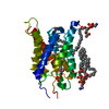
Mass: 744.034 Da / Num. of mol.: 5 / Source method: obtained synthetically / Formula: C41H78NO8P / Comment: DOPE, phospholipid*YM
#3: Water ChemComp-HOH / water / Water


Mass: 18.015 Da / Num. of mol.: 14 / Source method: isolated from a natural source / Formula: H2O

-
Experimental details

-
Experiment

ExperimentMethod: ELECTRON CRYSTALLOGRAPHY / Number of used crystals: 1
EM experimentAggregation state: 2D ARRAY / 3D reconstruction method: electron crystallography

-
Sample preparation

ComponentName: aquaporin-4 S180D mutant / Type: COMPLEX
SpecimenEmbedding applied: NO / Shadowing applied: NO / Staining applied: NO / Vitrification applied: YES
Crystal growTemperature: 293 K / Method: microdialysis / pH: 6
Details: 10mM MES (pH 6.0), 75mM NaCl, 50mM MgCl2, 2mM DTT, 1% glycerol, MICRODIALYSIS, temperature 293K

-
Data collection

MicroscopyModel: JEOL 3000SFF
Electron gunElectron source: FIELD EMISSION GUN / Accelerating voltage: 300 kV / Illumination mode: FLOOD BEAM
Electron lensMode: DIFFRACTION
Image recordingFilm or detector model: GENERIC CCD
DiffractionMean temperature: 4.2 K
Diffraction sourceSource: ELECTRON MICROSCOPE / Type: OTHER / Wavelength: 2.8 Å
DetectorType: Tietz / Detector: CCD / Date: Feb 4, 2008
RadiationProtocol: SINGLE WAVELENGTH / Monochromatic (M) / Laue (L): M / Scattering type: electron
Radiation wavelengthWavelength: 2.8 Å / Relative weight: 1
ReflectionResolution: 2.8→16.89 Å / Num. all: 10057 / Num. obs: 8750 / % possible obs: 87 % / Observed criterion σ(F): 0 / Observed criterion σ(I): 0 / Rmerge(I) obs: 0.197 / Rsym value: 0.197
Reflection shellResolution: 2.8→2.87 Å / % possible all: 80.15

-
Processing

Software
NameVersionClassification
REFMAC5.2.0019refinement
MRCdata collection
MRCdata reduction
MRCdata scaling
CNSphasing
3D reconstructionSymmetry type: 2D CRYSTAL
RefinementMethod to determine structure: MOLECULAR REPLACEMENT
Starting model: 2D57
Resolution: 2.8→10 Å / Cor.coef. Fo:Fc: 0.926 / Cor.coef. Fo:Fc free: 0.839 / SU B: 41.271 / SU ML: 0.355 / Cross valid method: THROUGHOUT / σ(F): 0 / σ(I): 0 / ESU R Free: 0.395 / Stereochemistry target values: MAXIMUM LIKELIHOOD / Details: HYDROGENS HAVE BEEN ADDED IN THE RIDING POSITIONS
RfactorNum. reflection% reflectionSelection details
Rfree0.28788 481 4.9 %RANDOM
Rwork0.23079 ---
all0.234 9879 --
obs0.234 8089 81.88 %-
Solvent computationIon probe radii: 0.8 Å / Shrinkage radii: 0.8 Å / VDW probe radii: 1.2 Å / Solvent model: MASK
Displacement parametersBiso mean: 52.335 Å2
Baniso -1Baniso -2Baniso -3
1--6.68 Å20 Å20 Å2
2---6.68 Å20 Å2
3---13.37 Å2
Refinement stepCycle: LAST / Resolution: 2.8→10 Å
ProteinNucleic acidLigandSolventTotal
Num. atoms1642 0 211 14 1867
Refine LS restraints
Refine-IDTypeDev idealDev ideal targetNumber
ELECTRON CRYSTALLOGRAPHYr_bond_refined_d0.0110.0221889
ELECTRON CRYSTALLOGRAPHYr_angle_refined_deg1.5242.0272522
ELECTRON CRYSTALLOGRAPHYr_dihedral_angle_1_deg6.2975221
ELECTRON CRYSTALLOGRAPHYr_dihedral_angle_2_deg31.61523.01953
ELECTRON CRYSTALLOGRAPHYr_dihedral_angle_3_deg21.86815244
ELECTRON CRYSTALLOGRAPHYr_dihedral_angle_4_deg18.107153
ELECTRON CRYSTALLOGRAPHYr_chiral_restr0.090.2279
ELECTRON CRYSTALLOGRAPHYr_gen_planes_refined0.0040.021282
ELECTRON CRYSTALLOGRAPHYr_nbd_refined0.2610.21154
ELECTRON CRYSTALLOGRAPHYr_nbtor_refined0.3210.21286
ELECTRON CRYSTALLOGRAPHYr_xyhbond_nbd_refined0.1790.272
ELECTRON CRYSTALLOGRAPHYr_symmetry_vdw_refined0.2510.2107
ELECTRON CRYSTALLOGRAPHYr_symmetry_hbond_refined0.1020.23
ELECTRON CRYSTALLOGRAPHYr_mcbond_it0.6531.51113
ELECTRON CRYSTALLOGRAPHYr_mcangle_it1.15421749
ELECTRON CRYSTALLOGRAPHYr_scbond_it0.9153869
ELECTRON CRYSTALLOGRAPHYr_scangle_it1.5324.5773
ELECTRON CRYSTALLOGRAPHYr_rigid_bond_restr0.73131982
ELECTRON CRYSTALLOGRAPHYr_sphericity_free4.147314
ELECTRON CRYSTALLOGRAPHYr_sphericity_bonded0.77731853
LS refinement shellResolution: 2.8→2.867 Å / Total num. of bins used: 20
RfactorNum. reflection% reflection
Rfree0.326 31 -
Rwork0.337 518 -
obs--80.15 %

+
About Yorodumi

-
News

-
Feb 9, 2022. New format data for meta-information of EMDB entries

New format data for meta-information of EMDB entries

  • Version 3 of the EMDB header file is now the official format.
  • The previous official version 1.9 will be removed from the archive.

Related info.:EMDB header

External links:wwPDB to switch to version 3 of the EMDB data model

-
Aug 12, 2020. Covid-19 info

Covid-19 info

URL: https://pdbj.org/emnavi/covid19.php

New page: Covid-19 featured information page in EM Navigator.

Related info.:Covid-19 info / Mar 5, 2020. Novel coronavirus structure data

+
Mar 5, 2020. Novel coronavirus structure data

Novel coronavirus structure data

Related info.:Yorodumi Speices / Aug 12, 2020. Covid-19 info

External links:COVID-19 featured content - PDBj / Molecule of the Month (242):Coronavirus Proteases

+
Jan 31, 2019. EMDB accession codes are about to change! (news from PDBe EMDB page)

EMDB accession codes are about to change! (news from PDBe EMDB page)

  • The allocation of 4 digits for EMDB accession codes will soon come to an end. Whilst these codes will remain in use, new EMDB accession codes will include an additional digit and will expand incrementally as the available range of codes is exhausted. The current 4-digit format prefixed with “EMD-” (i.e. EMD-XXXX) will advance to a 5-digit format (i.e. EMD-XXXXX), and so on. It is currently estimated that the 4-digit codes will be depleted around Spring 2019, at which point the 5-digit format will come into force.
  • The EM Navigator/Yorodumi systems omit the EMD- prefix.

Related info.:Q: What is EMD? / ID/Accession-code notation in Yorodumi/EM Navigator

External links:EMDB Accession Codes are Changing Soon! / Contact to PDBj

+
Jul 12, 2017. Major update of PDB

Major update of PDB

  • wwPDB released updated PDB data conforming to the new PDBx/mmCIF dictionary.
  • This is a major update changing the version number from 4 to 5, and with Remediation, in which all the entries are updated.
  • In this update, many items about electron microscopy experimental information are reorganized (e.g. em_software).
  • Now, EM Navigator and Yorodumi are based on the updated data.

External links:wwPDB Remediation / Enriched Model Files Conforming to OneDep Data Standards Now Available in the PDB FTP Archive

-
Yorodumi

Thousand views of thousand structures

  • Yorodumi is a browser for structure data from EMDB, PDB, SASBDB, etc.
  • This page is also the successor to EM Navigator detail page, and also detail information page/front-end page for Omokage search.
  • The word "yorodu" (or yorozu) is an old Japanese word meaning "ten thousand". "mi" (miru) is to see.

Related info.:EMDB / PDB / SASBDB / Comparison of 3 databanks / Yorodumi Search / Aug 31, 2016. New EM Navigator & Yorodumi / Yorodumi Papers / Jmol/JSmol / Function and homology information / Changes in new EM Navigator and Yorodumi

Read more