 Movie
Movie Controller


+ Open data

- Basic information
| Entry | Database: PDB / ID: 2x7n | ||||||
|---|---|---|---|---|---|---|---|
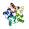
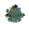
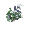
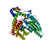
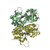
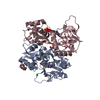
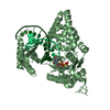
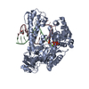
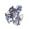
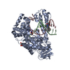
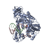
| Title | Mechanism of eIF6s anti-association activity | ||||||
 Components Components |
| ||||||
 Keywords Keywords |  RIBOSOMAL PROTEIN/RNA / RIBOSOMAL PROTEIN-RNA COMPLEX / INITIATION FACTOR / PROTEIN BIOSYNTHESIS / RIBOSOMAL PROTEIN / RIBONUCLEOPROTEIN RIBOSOMAL PROTEIN/RNA / RIBOSOMAL PROTEIN-RNA COMPLEX / INITIATION FACTOR / PROTEIN BIOSYNTHESIS / RIBOSOMAL PROTEIN / RIBONUCLEOPROTEIN | ||||||
| Function / homology |  Function and homology information Function and homology informationmaturation of 5.8S rRNA from tricistronic rRNA transcript (SSU-rRNA, 5.8S rRNA, LSU-rRNA) / maturation of 5.8S rRNA / ribosomal subunit export from nucleus / SRP-dependent cotranslational protein targeting to membrane / GTP hydrolysis and joining of the 60S ribosomal subunit / Formation of a pool of free 40S subunits / ribosomal large subunit binding / Nonsense Mediated Decay (NMD) independent of the Exon Junction Complex (EJC) / Nonsense Mediated Decay (NMD) enhanced by the Exon Junction Complex (EJC) / preribosome, large subunit precursor ...maturation of 5.8S rRNA from tricistronic rRNA transcript (SSU-rRNA, 5.8S rRNA, LSU-rRNA) / maturation of 5.8S rRNA / ribosomal subunit export from nucleus / SRP-dependent cotranslational protein targeting to membrane / GTP hydrolysis and joining of the 60S ribosomal subunit / Formation of a pool of free 40S subunits / ribosomal large subunit binding / Nonsense Mediated Decay (NMD) independent of the Exon Junction Complex (EJC) / Nonsense Mediated Decay (NMD) enhanced by the Exon Junction Complex (EJC) / preribosome, large subunit precursor / L13a-mediated translational silencing of Ceruloplasmin expression / maturation of LSU-rRNA / ribosomal large subunit biogenesis / maturation of LSU-rRNA from tricistronic rRNA transcript (SSU-rRNA, 5.8S rRNA, LSU-rRNA) / translation initiation factor activity / assembly of large subunit precursor of preribosome / cytosolic ribosome assembly / rRNA processing / large ribosomal subunit rRNA binding / cytoplasmic translation / cytosolic large ribosomal subunit / structural constituent of ribosome / mRNA binding / nucleolus / RNA binding / nucleus / cytosol / cytoplasmSimilarity search - Function | ||||||
| Biological species |  SACCHAROMYCES CEREVISIAE (brewer's yeast) SACCHAROMYCES CEREVISIAE (brewer's yeast) | ||||||
| Method | ELECTRON MICROSCOPY / single particle reconstruction / cryo EM / Resolution: 11.8 Å | ||||||
 Authors Authors | Gartmann, M. / Blau, M. / Armache, J.-P. / Mielke, T. / Topf, M. / Beckmann, R. | ||||||
 Citation Citation | Journal: J Biol Chem / Year: 2010 Title: Mechanism of eIF6-mediated inhibition of ribosomal subunit joining. Authors: Marco Gartmann / Michael Blau / Jean-Paul Armache / Thorsten Mielke / Maya Topf / Roland Beckmann /   Abstract: During the process of ribosomal assembly, the essential eukaryotic translation initiation factor 6 (eIF6) is known to act as a ribosomal anti-association factor. However, a molecular understanding of ...During the process of ribosomal assembly, the essential eukaryotic translation initiation factor 6 (eIF6) is known to act as a ribosomal anti-association factor. However, a molecular understanding of the anti-association activity of eIF6 is still missing. Here we present the cryo-electron microscopy reconstruction of a complex of the large ribosomal subunit with eukaryotic eIF6 from Saccharomyces cerevisiae. The structure reveals that the eIF6 binding site involves mainly rpL23 (L14p in Escherichia coli). Based on our structural data, we propose that the mechanism of the anti-association activity of eIF6 is based on steric hindrance of intersubunit bridge formation around the dynamic bridge B6. | ||||||
| History |
| ||||||
| Remark 700 | SHEET DETERMINATION METHOD: DSSP THE SHEETS PRESENTED AS "CC" IN EACH CHAIN ON SHEET RECORDS BELOW ... SHEET DETERMINATION METHOD: DSSP THE SHEETS PRESENTED AS "CC" IN EACH CHAIN ON SHEET RECORDS BELOW IS ACTUALLY AN 5-STRANDED BARREL THIS IS REPRESENTED BY A 6-STRANDED SHEET IN WHICH THE FIRST AND LAST STRANDS ARE IDENTICAL. |
- Structure visualization
| Movie |
Movie viewer |
|---|---|
| Structure viewer | Molecule: MolmilJmol/JSmol |
- Downloads & links
-Download
| PDBx/mmCIF format | 2x7n.cif.gz | 100.7 KB | Display | PDBx/mmCIF format |
|---|---|---|---|---|
| PDB format | pdb2x7n.ent.gz | 78.4 KB | Display | PDB format |
| PDBx/mmJSON format | 2x7n.json.gz | Tree view | PDBx/mmJSON format | |
| Others |  Other downloads Other downloads |
-Validation report
| Arichive directory | https://data.pdbj.org/pub/pdb/validation_reports/x7/2x7nftp://data.pdbj.org/pub/pdb/validation_reports/x7/2x7n | HTTPS FTP |
|---|
-Related structure data
| Related structure data | 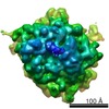 1705MC M: map data used to model this data C: citing same article ( |
|---|---|
| Similar structure data |


-Links
 PDBj
PDBj


- Assembly
| Deposited unit | 
|
|---|---|
| 1 |
|
-Components
| #1: RNA chain | Mass: 9042.445 Da / Num. of mol.: 1 / Fragment: 2684-2711 / Source method: isolated from a natural source / Source: (natural) SACCHAROMYCES CEREVISIAE (brewer's yeast) |
|---|---|
| #2: Protein | Mass: 24156.150 Da / Num. of mol.: 1 / Fragment: RESIDUES 1-224 / Source method: isolated from a natural source / Source: (natural) SACCHAROMYCES CEREVISIAE (brewer's yeast) / References: UniProt: Q12522 |
| #3: Protein | / YL32 / L17A Mass: 14047.472 Da / Num. of mol.: 1 / Fragment: RESIDUES 6-137 / Source method: isolated from a natural source / Source: (natural) SACCHAROMYCES CEREVISIAE (brewer's yeast) / References: UniProt: P04451, UniProt: P0CX41*PLUS |
| #4: Protein | Ribosome / L30 / YL221 / RP29 Mass: 6550.665 Da / Num. of mol.: 1 / Fragment: RESIDUES 1-56 / Source method: isolated from a natural source / Source: (natural) SACCHAROMYCES CEREVISIAE (brewer's yeast) / References: UniProt: P04449 |
-Experimental details
-Experiment
| Experiment | Method: ELECTRON MICROSCOPY |
|---|---|
| EM experiment | Aggregation state: PARTICLE / 3D reconstruction method: single particle reconstruction |
- Sample preparation
| Component | Name: 60S-EIF6 COMPLEX / Type: RIBOSOME / Details: CRYO-EM SINGLE-PARTICLE RECONSTRUCTION |
|---|---|
| Specimen | Embedding applied: NO / Shadowing applied: NO / Staining applied: NO / Vitrification applied: YES |
| Specimen support | Details: CARBON |
| Vitrification | Cryogen name: ETHANE / Details: ETHANE |
- Electron microscopy imaging
| Experimental equipment |  Model: Tecnai F30 / Image courtesy: FEI Company |
|---|---|
| Microscopy | Model: FEI TECNAI F30 |
| Electron gun | Electron source: FIELD EMISSION GUN / Accelerating voltage: 300 kV / Illumination mode: FLOOD BEAM |
| Electron lens | Mode: BRIGHT FIELDBright-field microscopy / Nominal magnification: 39000 X / Calibrated magnification: 38900 X / Cs: 2.26 mm |
| Specimen holder | Temperature: 95 K |
| Image recording | Electron dose: 20 e/Å2 / Film or detector model: KODAK SO-163 FILM |
| Radiation wavelength | Relative weight: 1 |
- Processing
| EM software | Name: SPIDER / Category: 3D reconstruction | ||||||||||||
|---|---|---|---|---|---|---|---|---|---|---|---|---|---|
| CTF correction | Details: DEFOCUS GROUP VOLUMES | ||||||||||||
| Symmetry | Point symmetry: C1 (asymmetric) | ||||||||||||
| 3D reconstruction | Resolution: 11.8 Å Details: SUBMISSION BASED ON EXPERIMENTAL DATA FROM EMDB EMD-1705. Symmetry type: POINT | ||||||||||||
| Atomic model building | Protocol: FLEXIBLE FIT / Space: REAL / Details: METHOD--FLEX-EM | ||||||||||||
| Atomic model building | PDB-ID: 1G62 | ||||||||||||
| Refinement | Highest resolution: 11.8 Å | ||||||||||||
| Refinement step | Cycle: LAST / Highest resolution: 11.8 Å
|
