 Movie
Movie Controller

[English] 日本語
 Yorodumi
- PDB-2r1g: Coordinates of the thermus thermophilus 30S components neighborin... -
+ Open data

- Basic information
| Entry | Database: PDB / ID: 2r1g | ||||||
|---|---|---|---|---|---|---|---|
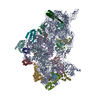
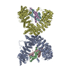
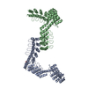
| Title | Coordinates of the thermus thermophilus 30S components neighboring RbfA as obtained by fitting into the CRYO-EM map of A 30S-RBFA complex | ||||||
 Components Components |
| ||||||
 Keywords Keywords |  RIBOSOMAL PROTEIN/RNA / 30S RIBOSOME MATURATION PROTEIN RbfA / COLD SHOCK RESPONSE PROTEIN RbfA / 30S-RbfA COMPLEX / RbfA BINDING SITE ON THE 30S / Ribonucleoprotein / Ribosomal protein / RNA-binding / rRNA-binding / tRNA-binding / Antibiotic resistance / RIBOSOMAL PROTEIN-RNA COMPLEX RIBOSOMAL PROTEIN/RNA / 30S RIBOSOME MATURATION PROTEIN RbfA / COLD SHOCK RESPONSE PROTEIN RbfA / 30S-RbfA COMPLEX / RbfA BINDING SITE ON THE 30S / Ribonucleoprotein / Ribosomal protein / RNA-binding / rRNA-binding / tRNA-binding / Antibiotic resistance / RIBOSOMAL PROTEIN-RNA COMPLEX | ||||||
| Function / homology |  Function and homology information Function and homology informationsmall ribosomal subunit / tRNA binding / rRNA binding / ribosome / structural constituent of ribosome / translation / ribonucleoprotein complex / response to antibiotic / cytoplasmSimilarity search - Function | ||||||
| Biological species |   Thermus thermophilus (bacteria) Thermus thermophilus (bacteria)synthetic construct (others) | ||||||
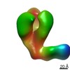
| Method | ELECTRON MICROSCOPY / single particle reconstruction / cryo EM / Resolution: 12.5 Å | ||||||
 Authors Authors | Datta, P.P. / Wilson, D.N. / Kawazoe, M. / Swami, N.K. / Kaminishi, T. / Sharma, M.R. / Booth, T.M. / Takemoto, C. / Fucini, P. / Yokoyama, S. / Agrawal, R.K. | ||||||
 Citation Citation | Journal: Mol Cell / Year: 2007 Title: Structural aspects of RbfA action during small ribosomal subunit assembly. Authors: Partha P Datta / Daniel N Wilson / Masahito Kawazoe / Neil K Swami / Tatsuya Kaminishi / Manjuli R Sharma / Timothy M Booth / Chie Takemoto / Paola Fucini / Shigeyuki Yokoyama / Rajendra K Agrawal /  Abstract: Ribosome binding factor A (RbfA) is a bacterial cold shock response protein, required for an efficient processing of the 5' end of the 16S ribosomal RNA (rRNA) during assembly of the small (30S) ...Ribosome binding factor A (RbfA) is a bacterial cold shock response protein, required for an efficient processing of the 5' end of the 16S ribosomal RNA (rRNA) during assembly of the small (30S) ribosomal subunit. Here we present a crystal structure of Thermus thermophilus (Tth) RbfA and a three-dimensional cryo-electron microscopic (EM) map of the Tth 30S*RbfA complex. RbfA binds to the 30S subunit in a position overlapping the binding sites of the A and P site tRNAs, and RbfA's functionally important C terminus extends toward the 5' end of the 16S rRNA. In the presence of RbfA, a portion of the 16S rRNA encompassing helix 44, which is known to be directly involved in mRNA decoding and tRNA binding, is displaced. These results shed light on the role played by RbfA during maturation of the 30S subunit, and also indicate how RbfA provides cells with a translational advantage under conditions of cold shock. #1: Journal: Nature / Year: 2000 Title: Molecular biology. Small subunit, big science. | ||||||
| History |
|
- Structure visualization
| Movie |
Movie viewer |
|---|---|
| Structure viewer | Molecule: MolmilJmol/JSmol |
- Downloads & links
-Download
| PDBx/mmCIF format | 2r1g.cif.gz | 35.7 KB | Display | PDBx/mmCIF format |
|---|---|---|---|---|
| PDB format | pdb2r1g.ent.gz | 16.9 KB | Display | PDB format |
| PDBx/mmJSON format | 2r1g.json.gz | Tree view | PDBx/mmJSON format | |
| Others |  Other downloads Other downloads |
-Validation report
| Arichive directory | https://data.pdbj.org/pub/pdb/validation_reports/r1/2r1gftp://data.pdbj.org/pub/pdb/validation_reports/r1/2r1g | HTTPS FTP |
|---|
-Related structure data
| Related structure data | 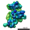 1413MC 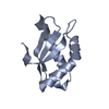 2dyjC  2r1cC C: citing same article ( M: map data used to model this data |
|---|---|
| Similar structure data |


-Links
 PDBj
PDBj


- Assembly
| Deposited unit | 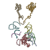
|
|---|---|
| 1 |
|
-Components
-16S RIBOSOMAL RNA HELIX ... , 7 types, 7 molecules ABCDEXF
| #1: RNA chain | Mass: 7781.646 Da / Num. of mol.: 1 / Source method: obtained synthetically / Source: (synth.) synthetic construct (others) |
|---|---|
| #2: RNA chain | Mass: 15544.350 Da / Num. of mol.: 1 / Source method: obtained synthetically / Source: (synth.) synthetic construct (others) |
| #3: RNA chain | Mass: 9440.723 Da / Num. of mol.: 1 / Source method: obtained synthetically / Source: (synth.) synthetic construct (others) |
| #4: RNA chain | Mass: 8969.390 Da / Num. of mol.: 1 / Source method: obtained synthetically / Source: (synth.) synthetic construct (others) |
| #5: RNA chain | Mass: 3457.122 Da / Num. of mol.: 1 / Source method: obtained synthetically / Source: (synth.) synthetic construct (others) |
| #6: RNA chain | Mass: 3530.178 Da / Num. of mol.: 1 / Source method: obtained synthetically / Source: (synth.) synthetic construct (others) |
| #7: RNA chain | Mass: 9434.667 Da / Num. of mol.: 1 / Source method: obtained synthetically / Source: (synth.) synthetic construct (others) |
-30S ribosomal protein ... , 3 types, 3 molecules GHI
| #8: Protein | / Coordinate model: Cα atoms only Mass: 14298.466 Da / Num. of mol.: 1 Source method: isolated from a genetically manipulated source Source: (gene. exp.) Thermus thermophilus (bacteria) / Strain: HB27 / Gene: rpsI, rps9 / Production host: Escherichia coli (E. coli) / References: UniProt: P62669 |
|---|---|
| #9: Protein | / Coordinate model: Cα atoms only Mass: 13804.311 Da / Num. of mol.: 1 / Fragment: residues 5-128 Source method: isolated from a genetically manipulated source Source: (gene. exp.) Thermus thermophilus (bacteria) / Strain: HB27 / Gene: rpsL, rps12 / Production host: Escherichia coli (E. coli) / References: UniProt: P17293, UniProt: P61941*PLUS |
| #10: Protein | / Coordinate model: Cα atoms only Mass: 14207.666 Da / Num. of mol.: 1 Source method: isolated from a genetically manipulated source Source: (gene. exp.) Thermus thermophilus (bacteria) / Strain: HB27 / Gene: rpsM, rps13 / Production host: Escherichia coli (E. coli) / References: UniProt: P62655 |
-Details
| Sequence details | SICNE THE PORTION (1411-1489) OF H44 IS NOT CLOSE TO THE RBFA, THE AUTHOR DID NOT PROVIDE THOSE ...SICNE THE PORTION (1411-1489) OF H44 IS NOT CLOSE TO THE RBFA, THE AUTHOR DID NOT PROVIDE THOSE COORDINATE |
|---|
-Experimental details
-Experiment
| Experiment | Method: ELECTRON MICROSCOPY |
|---|---|
| EM experiment | Aggregation state: PARTICLE / 3D reconstruction method: single particle reconstruction |
- Sample preparation
| Component | Name: THERMUS THERMOPHILUS 30S RIBOSOMAL SUBUNIT COMPLEXED WITH RBFA Type: RIBOSOME / Details: RBFA WAS BOUND TO S1-DEPLETED 30S SUBUNIT |
|---|---|
| Buffer solution | Name: 20mM, Hepes-KOH (pH 7.8), 10mM Mg(OAc)2, 200mM NH4Cl, 65mM KCl pH: 7.8 Details: 20mM, Hepes-KOH (pH 7.8), 10mM Mg(OAc)2, 200mM NH4Cl, 65mM KCl |
| Specimen | Conc.: 0.03 mg/ml / Embedding applied: NO / Shadowing applied: NO / Staining applied: NO / Vitrification applied: YES |
| Specimen support | Details: QUANTIFOIL HOLEY-CRBON FILM GRID |
| Vitrification | Instrument: HOMEMADE PLUNGER / Cryogen name: ETHANE / Details: RAPID-FREEZING IN LIQUID ETHANE |
- Electron microscopy imaging
| Experimental equipment |  Model: Tecnai F20 / Image courtesy: FEI Company |
|---|---|
| Microscopy | Model: FEI TECNAI F20 / Date: Jan 18, 2005 / Details: ZEISS IMAGING SCANNER, STEP SIZE 14micro-m |
| Electron gun | Electron source: FIELD EMISSION GUN / Accelerating voltage: 200 kV / Illumination mode: FLOOD BEAM |
| Electron lens | Mode: BRIGHT FIELDBright-field microscopy / Nominal magnification: 50000 X / Calibrated magnification: 50760 X / Nominal defocus max: 3500 nm / Nominal defocus min: 700 nm / Cs: 2 mm |
| Specimen holder | Temperature: 93 K / Tilt angle max: 0 ° / Tilt angle min: 0 ° |
| Image recording | Electron dose: 20 e/Å2 / Film or detector model: KODAK SO-163 FILM |
| Image scans | Num. digital images: 131 |
| Radiation | Protocol: SINGLE WAVELENGTH / Monochromatic (M) / Laue (L): M / Scattering type: x-ray |
| Radiation wavelength | Relative weight: 1 |
- Processing
| EM software |
| ||||||||||||
|---|---|---|---|---|---|---|---|---|---|---|---|---|---|
| CTF correction | Details: CTF CORRECTION OF 3D-MAPS BY WIENER FILTRATION | ||||||||||||
| Symmetry | Point symmetry: C1 (asymmetric) | ||||||||||||
| 3D reconstruction | Method: CRYO-ELECTRON MICROSCOPY AND 3D IMAGE PROCESSING / Resolution: 12.5 Å / Num. of particles: 61207 / Actual pixel size: 2.76 Å / Magnification calibration: TMV Details: This entry contains only a CA trace for the protein and only phosphorus atom for the RNA in the coordinate. CROSS-CORRELATION COEFFICIENT (CCF) VALUE FOR RBFA HOMOLOGY MODEL FITTED INTO THE ...Details: This entry contains only a CA trace for the protein and only phosphorus atom for the RNA in the coordinate. CROSS-CORRELATION COEFFICIENT (CCF) VALUE FOR RBFA HOMOLOGY MODEL FITTED INTO THE CORRESPONDING CRYO-EM DENSITY WAS 0.79 Symmetry type: POINT | ||||||||||||
| Atomic model building | Protocol: RIGID BODY FIT / Space: REAL Target criteria: X-RAY COORDINATES OF T. THERMOPHILUS 30S RIBOSOMAL SUBUNIT AND THE HOMOLOGY MODEL OF T. THERMOPHILUS RBFA WERE FITTED INTO THE 12.5 ANGSTROMS RESOLUTION CRYO-EM MAP OF THE T. ...Target criteria: X-RAY COORDINATES OF T. THERMOPHILUS 30S RIBOSOMAL SUBUNIT AND THE HOMOLOGY MODEL OF T. THERMOPHILUS RBFA WERE FITTED INTO THE 12.5 ANGSTROMS RESOLUTION CRYO-EM MAP OF THE T. THERMOPHILUS 30S SUBUNIT-RBFA COMPLEX. ALL THE ATOMIC COORDINATES WERE FITTED AS RIGID BODIES Details: METHOD--CROSS-CORRELATION BASED MANUAL FITTING IN O REFINEMENT PROTOCOL--MULTIPLE RIGID BODY | ||||||||||||
| Atomic model building | PDB-ID: 1J5E Accession code: 1J5E / Source name: PDB / Type: experimental model | ||||||||||||
| Refinement step | Cycle: LAST
|
