Movie
Movie Controller


+ Open data

- Basic information
| Entry | Database: PDB / ID: 2iy3 | ||||||
|---|---|---|---|---|---|---|---|
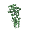
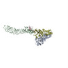
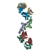
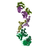
| Title | Structure of the E. Coli Signal Regognition Particle | ||||||
 Components Components |
| ||||||
 Keywords Keywords | RNA-BINDING / RNA-BINDING PROTEIN COMPLEX /  SIGNAL RECOGNITION PARTICLE SIGNAL RECOGNITION PARTICLE | ||||||
| Function / homology |  Function and homology informationsignal recognition particle / signal-recognition-particle GTPase / 7S RNA binding / SRP-dependent cotranslational protein targeting to membrane / GTPase activity / GTP binding / ATP hydrolysis activity Function and homology informationsignal recognition particle / signal-recognition-particle GTPase / 7S RNA binding / SRP-dependent cotranslational protein targeting to membrane / GTPase activity / GTP binding / ATP hydrolysis activitySimilarity search - Function | ||||||
| Biological species |   Thermus aquaticus (bacteria) Thermus aquaticus (bacteria) Sulfolobus solfataricus (archaea)Escherichia coli (E. coli) Sulfolobus solfataricus (archaea)Escherichia coli (E. coli)synthetic construct (others) | ||||||
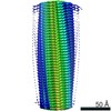
| Method | ELECTRON MICROSCOPY / single particle reconstruction / cryo EM / Resolution: 16 Å | ||||||
| Model type details | CA ATOMS ONLY, CHAIN A, C ; P ATOMS ONLY, CHAIN B | ||||||
 Authors Authors | Schaffitzel, C. / Oswald, M. / Berger, I. / Ishikawa, T. / Abrahams, J.P. / Koerten, H.K. / Koning, R.I. / Ban, N. | ||||||
 Citation Citation | Journal: Nature / Year: 2006 Title: Structure of the E. coli signal recognition particle bound to a translating ribosome. Authors: Christiane Schaffitzel / Miro Oswald / Imre Berger / Takashi Ishikawa / Jan Pieter Abrahams / Henk K Koerten / Roman I Koning / Nenad Ban /  Abstract: The prokaryotic signal recognition particle (SRP) targets membrane proteins into the inner membrane. It binds translating ribosomes and screens the emerging nascent chain for a hydrophobic signal ...The prokaryotic signal recognition particle (SRP) targets membrane proteins into the inner membrane. It binds translating ribosomes and screens the emerging nascent chain for a hydrophobic signal sequence, such as the transmembrane helix of inner membrane proteins. If such a sequence emerges, the SRP binds tightly, allowing the SRP receptor to lock on. This assembly delivers the ribosome-nascent chain complex to the protein translocation machinery in the membrane. Using cryo-electron microscopy and single-particle reconstruction, we obtained a 16 A structure of the Escherichia coli SRP in complex with a translating E. coli ribosome containing a nascent chain with a transmembrane helix anchor. We also obtained structural information on the SRP bound to an empty E. coli ribosome. The latter might share characteristics with a scanning SRP complex, whereas the former represents the next step: the targeting complex ready for receptor binding. High-resolution structures of the bacterial ribosome and of the bacterial SRP components are available, and their fitting explains our electron microscopic density. The structures reveal the regions that are involved in complex formation, provide insight into the conformation of the SRP on the ribosome and indicate the conformational changes that accompany high-affinity SRP binding to ribosome nascent chain complexes upon recognition of the signal sequence. | ||||||
| History |
|
- Structure visualization
| Movie |
Movie viewer |
|---|---|
| Structure viewer | Molecule: MolmilJmol/JSmol |
- Downloads & links
-Download
| PDBx/mmCIF format | 2iy3.cif.gz | 27.2 KB | Display | PDBx/mmCIF format |
|---|---|---|---|---|
| PDB format | pdb2iy3.ent.gz | 15.1 KB | Display | PDB format |
| PDBx/mmJSON format | 2iy3.json.gz | Tree view | PDBx/mmJSON format | |
| Others |  Other downloads Other downloads |
-Validation report
| Arichive directory | https://data.pdbj.org/pub/pdb/validation_reports/iy/2iy3ftp://data.pdbj.org/pub/pdb/validation_reports/iy/2iy3 | HTTPS FTP |
|---|
-Related structure data
| Related structure data |  1250MC 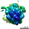 1251MC M: map data used to model this data C: citing same article ( |
|---|---|
| Similar structure data |


-Links
 PDBj
PDBj


- Assembly
| Deposited unit | 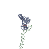
|
|---|---|
| 1 |
|
-Components
| #1: Protein | / Fifty-four homolog / SRP54 / Coordinate model: Cα atoms only Mass: 48274.094 Da / Num. of mol.: 1 Source method: isolated from a genetically manipulated source Source: (gene. exp.) Thermus aquaticus (bacteria), (gene. exp.) Sulfolobus solfataricus (archaea)Gene: ffh, srp54, SULA_1982, SULB_1983, SULC_1981 / Plasmid: PET24A_FFH / Production host: Escherichia coli (E. coli) / Strain (production host): BL21(DE3) / References: UniProt: O07347, UniProt: A0A0E3MG81 |
|---|---|
| #2: RNA chain | Mass: 35547.090 Da / Num. of mol.: 1 / Source method: obtained synthetically / Source: (synth.) Escherichia coli (E. coli) / References: GenBank: 1126835767 |
| #3: Protein/peptide | / Coordinate model: Cα atoms only Mass: 1380.632 Da / Num. of mol.: 1 / Source method: obtained synthetically / Source: (synth.) synthetic construct |
-Experimental details
-Experiment
| Experiment | Method: ELECTRON MICROSCOPY |
|---|---|
| EM experiment | Aggregation state: PARTICLE / 3D reconstruction method: single particle reconstruction |
- Sample preparation
| Component | Name: SRP BOUND TO RIBOSOME / Type: RIBOSOME |
|---|---|
| Buffer solution | Name: 50 MM HEPES-KOH PH 7.5, 100 MM KCL, 25 MM MGCL2, 1 MM DTT,1 MM GTP pH: 7.5 Details: 50 MM HEPES-KOH PH 7.5, 100 MM KCL, 25 MM MGCL2, 1 MM DTT,1 MM GTP |
| Specimen | Embedding applied: NO / Shadowing applied: NO / Staining applied: NO / Vitrification applied: YES |
| Specimen support | Details: HOLEY CARBON |
| Vitrification | Instrument: HOMEMADE PLUNGER / Cryogen name: ETHANE / Details: PLUNGED INTO ETHANE |
- Electron microscopy imaging
| Microscopy | Model: FEI TECNAI 20 / Date: Jun 2, 2005 |
|---|---|
| Electron gun | Electron source: FIELD EMISSION GUN / Accelerating voltage: 200 kV / Illumination mode: FLOOD BEAM |
| Electron lens | Mode: BRIGHT FIELDBright-field microscopy / Nominal magnification: 50000 X / Calibrated magnification: 51000 X / Nominal defocus max: 4500 nm / Nominal defocus min: 1000 nm / Cs: 2 mm |
| Specimen holder | Temperature: 77 K / Tilt angle max: 0 ° / Tilt angle min: 0 ° |
| Image recording | Electron dose: 10 e/Å2 / Film or detector model: KODAK SO-163 FILM |
| Image scans | Num. digital images: 251 |
- Processing
| EM software |
| ||||||||||||
|---|---|---|---|---|---|---|---|---|---|---|---|---|---|
| CTF correction | Details: PHASE FLIPPING | ||||||||||||
| Symmetry | Point symmetry: C1 (asymmetric) | ||||||||||||
| 3D reconstruction | Method: PROJECTION MATCHING / Resolution: 16 Å / Num. of particles: 24382 / Nominal pixel size: 3.81 Å Details: THE COORDINATES BELOW ARE MADE UP FROM THE FOLLOWING COMPONENTS: FFH NG DOMAIN (RESIDUE 1-296), ORGANISM SCIENTIFIC: THERMUS AQUATICUS (PDB ENTRY 1JPN CHAIN A). FFH M DOMAIN (RESIDUE 297-432) ...Details: THE COORDINATES BELOW ARE MADE UP FROM THE FOLLOWING COMPONENTS: FFH NG DOMAIN (RESIDUE 1-296), ORGANISM SCIENTIFIC: THERMUS AQUATICUS (PDB ENTRY 1JPN CHAIN A). FFH M DOMAIN (RESIDUE 297-432), ORANISM SCIENTIFIC: SULFOLOBUS SOLFATARICUS (PDB ENTRY 1QZW CHAIN A). 4.5S RNA (RESIDUE 33- 73), ORGANISM SCIENTIFIC: SULFOLOBUS SOLFATARICUS (PDB ENTRY 1QZW, CHAIN B). 4.5S RNA (RESIDUE 1-32 AND 74-110), ORGANISM SCIENTIFIC: ESCHERICHIA COLI (PDB: SRPDB, ROSENBLAD ET AL, NUCLEIC ACID RES 21, 363-364,12:2003. Symmetry type: POINT | ||||||||||||
| Atomic model building | Protocol: OTHER / Space: REAL / Target criteria: Cross-correlation coefficient Details: METHOD--LOCAL CORRELATION REFINEMENT PROTOCOL--X-RAY | ||||||||||||
| Atomic model building |
| ||||||||||||
| Refinement | Highest resolution: 16 Å | ||||||||||||
| Refinement step | Cycle: LAST / Highest resolution: 16 Å
|