 Movie
Movie Controller

[English] 日本語
 Yorodumi
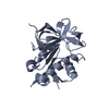
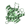
- PDB-2i68: Cryo-EM based theoretical model structure of transmembrane domain... -
+ Open data

- Basic information
| Entry | Database: PDB / ID: 2i68 | ||||||
|---|---|---|---|---|---|---|---|
| Title | Cryo-EM based theoretical model structure of transmembrane domain of the multidrug-resistance antiporter from E. coli EmrE | ||||||
 Components Components | Protein emrE | ||||||
 Keywords Keywords |  TRANSPORT PROTEIN / transmembrane protein / small-multidrug resistance / transporter / homodimer / dual topology TRANSPORT PROTEIN / transmembrane protein / small-multidrug resistance / transporter / homodimer / dual topology | ||||||
| Function / homology |  Function and homology information Function and homology informationEmrE multidrug transporter complex / amino-acid betaine transmembrane transporter activity / choline transmembrane transporter activity / glycine betaine transport / choline transport / xenobiotic detoxification by transmembrane export across the plasma membrane / xenobiotic transport / antiporter activity / response to osmotic stress / xenobiotic transmembrane transporter activity ...EmrE multidrug transporter complex / amino-acid betaine transmembrane transporter activity / choline transmembrane transporter activity / glycine betaine transport / choline transport / xenobiotic detoxification by transmembrane export across the plasma membrane / xenobiotic transport / antiporter activity / response to osmotic stress / xenobiotic transmembrane transporter activity / transmembrane transporter activity / xenobiotic metabolic process / transmembrane transport / cellular response to xenobiotic stimulus / response to xenobiotic stimulus / DNA damage response / membrane / identical protein binding / plasma membraneSimilarity search - Function | ||||||
| Biological species |  Escherichia coli (E. coli) Escherichia coli (E. coli) | ||||||
| Method | ELECTRON CRYSTALLOGRAPHY / electron crystallography / cryo EM / Resolution: 7.5 Å | ||||||
 Authors Authors | Fleishman, S.J. / Harrington, S.E. / Enosh, A. / Halperin, D. / Tate, C.G. / Ben-Tal, N. | ||||||
 Citation Citation | Journal: J Mol Biol / Year: 2006 Title: Quasi-symmetry in the cryo-EM structure of EmrE provides the key to modeling its transmembrane domain. Authors: Sarel J Fleishman / Susan E Harrington / Angela Enosh / Dan Halperin / Christopher G Tate / Nir Ben-Tal /  Abstract: Small multidrug resistance (SMR) transporters contribute to bacterial resistance by coupling the efflux of a wide range of toxic aromatic cations, some of which are commonly used as antibiotics and ...Small multidrug resistance (SMR) transporters contribute to bacterial resistance by coupling the efflux of a wide range of toxic aromatic cations, some of which are commonly used as antibiotics and antiseptics, to proton influx. EmrE is a prototypical small multidrug resistance transporter comprising four transmembrane segments (M1-M4) that forms dimers. It was suggested recently that EmrE molecules in the dimer have different topologies, i.e. monomers have opposite orientations with respect to the membrane plane. A 3-D structure of EmrE acquired by electron cryo-microscopy (cryo-EM) at 7.5 Angstroms resolution in the membrane plane showed that parts of the structure are related by quasi-symmetry. We used this symmetry relationship, combined with sequence conservation data, to assign the transmembrane segments in EmrE to the densities seen in the cryo-EM structure. A C alpha model of the transmembrane region was constructed by considering the evolutionary conservation pattern of each helix. The model is validated by much of the biochemical data on EmrE with most of the positions that were identified as affecting substrate translocation being located around the substrate-binding cavity. A suggested mechanism for proton-coupled substrate translocation in small multidrug resistance antiporters provides a mechanistic rationale to the experimentally observed inverted topology. #1: Journal: EMBO J / Year: 2003Title: Three-dimensional structure of the bacterial multidrug transporter EmrE shows it is an asymmetric homodimer. Authors: Iban Ubarretxena-Belandia / Joyce M Baldwin / Shimon Schuldiner / Christopher G Tate /  Abstract: The small multidrug resistance family of transporters is widespread in bacteria and is responsible for bacterial resistance to toxic aromatic cations by proton-linked efflux. We have determined the ...The small multidrug resistance family of transporters is widespread in bacteria and is responsible for bacterial resistance to toxic aromatic cations by proton-linked efflux. We have determined the three-dimensional (3D) structure of the Escherichia coli multidrug transporter EmrE by electron cryomicroscopy of 2D crystals, including data to 7.0 A resolution. The structure of EmrE consists of a bundle of eight transmembrane alpha-helices with one substrate molecule bound near the centre. The substrate binding chamber is formed from six helices and is accessible both from the aqueous phase and laterally from the lipid bilayer. The most remarkable feature of the structure of EmrE is that it is an asymmetric homodimer. The possible arrangement of the two polypeptides in the EmrE dimer is discussed based on the 3D density map. | ||||||
| History |
|
- Structure visualization
| Movie |
Movie viewer |
|---|---|
| Structure viewer | Molecule: MolmilJmol/JSmol |
- Downloads & links
-Download
| PDBx/mmCIF format | 2i68.cif.gz | 31.7 KB | Display | PDBx/mmCIF format |
|---|---|---|---|---|
| PDB format | pdb2i68.ent.gz | 15.1 KB | Display | PDB format |
| PDBx/mmJSON format | 2i68.json.gz | Tree view | PDBx/mmJSON format | |
| Others |  Other downloads Other downloads |
-Validation report
| Arichive directory | https://data.pdbj.org/pub/pdb/validation_reports/i6/2i68ftp://data.pdbj.org/pub/pdb/validation_reports/i6/2i68 | HTTPS FTP |
|---|
-Related structure data
| Related structure data | 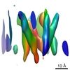 1087M M: map data used to model this data |
|---|---|
| Similar structure data |


-Links
 PDBj
PDBj- Assembly
| Deposited unit | 
| ||||||||
|---|---|---|---|---|---|---|---|---|---|
| 1 |
| ||||||||
| Unit cell |
|
-Components
| #1: Protein | Mass: 15203.710 Da / Num. of mol.: 2 Source method: isolated from a genetically manipulated source Source: (gene. exp.) Escherichia coli (E. coli) / Gene: emrE, EB, mvrC / Plasmid: T7-7 / Production host: Escherichia coli (E. coli) / Strain (production host): TA15(pGp1-2) / References: UniProt: P23895 |
|---|
-Experimental details
-Experiment
| Experiment | Method: ELECTRON CRYSTALLOGRAPHY |
|---|---|
| EM experiment | Aggregation state: 2D ARRAY / 3D reconstruction method: electron crystallography |
- Sample preparation
| Component | Name: multidrug-resistance antiporter from E. coli EmrE / Type: COMPLEX |
|---|---|
| Buffer solution | pH: 7.5 / Details: 20 mM Sodium phosphate pH7.5, 100 mM NaCl, 2mM |
| Specimen | Embedding applied: NO / Shadowing applied: NO / Staining applied: NO / Vitrification applied: YES |
| Vitrification | Instrument: HOMEMADE PLUNGER / Cryogen name: NITROGEN |
-Data collection
| Experimental equipment |  Model: Tecnai F30 / Image courtesy: FEI Company |
|---|---|
| Microscopy | Model: FEI TECNAI F30 / Date: Jan 1, 2003 |
| Electron gun | Electron source: FIELD EMISSION GUN / Accelerating voltage: 300 kV / Illumination mode: SPOT SCAN |
| Electron lens | Mode: BRIGHT FIELDBright-field microscopy / Nominal magnification: 60000 X / Nominal defocus max: 1600 nm / Nominal defocus min: 200 nm |
| Image recording | Electron dose: 15 e/Å2 / Film or detector model: KODAK SO-163 FILM |
| Radiation | Protocol: SINGLE WAVELENGTH / Monochromatic (M) / Laue (L): M / Scattering type: electron |
| Radiation wavelength | Relative weight: 1 |
- Processing
| 3D reconstruction | Resolution: 7.5 Å / Resolution method: OTHER Details: Canonical alpha-helices were fitted into a cryo-EM structure of EmrE at 6Angstroms in-plane and 16Angstroms vertical resolution. The sequence segments were assigned based on biophysical and ...Details: Canonical alpha-helices were fitted into a cryo-EM structure of EmrE at 6Angstroms in-plane and 16Angstroms vertical resolution. The sequence segments were assigned based on biophysical and sequence data as elaborated in the principal citation. The orientation of each helix around its principal axis was set using evolutionary conservation, requiring that evolutionarily conserved positions be packed inside the core of the protein, whereas variable residues face the outside. A kink was introduced in helix C to fit a bend in the cryo-EM structure and according to sequence clues (see principal citation). A full description of potential inaccuracies in the model is presented in the principal citation. In brief, these include the following: the vertical positioning of the helices may be wrong by several Angstroms due to the low vertical resolution of the cryo-EM structure; the orientations of the helices around their principal axes may vary by about 20 degrees; the positions of backbone atoms on the terminal turns of each helix may not conform to alpha-helical ideality as assumed in the model structure. Symmetry type: 2D CRYSTAL | ||||||||||||
|---|---|---|---|---|---|---|---|---|---|---|---|---|---|
| Refinement step | Cycle: LAST
|
