Journal: EMBO J / Year: 2006 Title: High-resolution cryo-EM maps show the nucleotide binding pocket of KIF1A in open and closed conformations. Authors: Masahide Kikkawa / Nobutaka Hirokawa / Abstract: Kinesin is an ATP-driven microtubule (MT)-based motor fundamental to organelle transport. Although a number of kinesin crystal structures have been solved, the structural evidence for coupling ...Kinesin is an ATP-driven microtubule (MT)-based motor fundamental to organelle transport. Although a number of kinesin crystal structures have been solved, the structural evidence for coupling between the bound nucleotide and the conformation of kinesin is elusive. In addition, the structural basis of the MT-induced ATPase activity of kinesin is not clear because of the absence of the MT in the structure. Here, we report cryo-electron microscopy structures of the monomeric kinesin KIF1A-MT complex in two nucleotide states at about 10 A resolution, sufficient to reveal the secondary structure. These high-resolution maps visualized clear structural changes that suggest a mechanical pathway from the nucleotide to the neck linker via the motor core rotation. In addition, new nucleotide binding pocket conformations are observed that are different from X-ray crystallographic structures; it is closed in the 5'-adenylyl-imidodiphosphate state, but open in the ADP state. These results suggest a structural model of biased diffusion movement of monomeric kinesin motor.
Aggregation state: FILAMENT / 3D reconstruction method: helical reconstruction
-
Sample preparation
Component
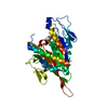
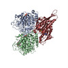
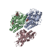
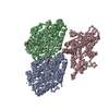
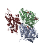
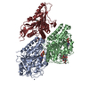
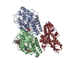
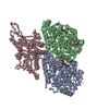
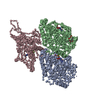
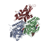
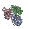
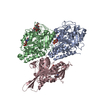
Name: KIF1A head decorated-microtubule complex / Type: COMPLEX Details: SAMPLE DETAILS: TUBULIN WAS POLYMERIZED IN PEM BUFFER (PIPES 100 MM, PH 6.8, EGTA 1 MM, MGCL2, GTP 1MM, PACLITAXEL 10 MICRO M, 5% DMSO ) FOR 2 HRS AT 37C. A DROP OF THE POLYMERIZED ...Details: SAMPLE DETAILS: TUBULIN WAS POLYMERIZED IN PEM BUFFER (PIPES 100 MM, PH 6.8, EGTA 1 MM, MGCL2, GTP 1MM, PACLITAXEL 10 MICRO M, 5% DMSO ) FOR 2 HRS AT 37C. A DROP OF THE POLYMERIZED MICROTUBULE WAS PLACED ON THE HOLEY CARBON FILM ON THE EM-GRID AND LEFT FOR 10 S IN THE HUMID CHAMBER. THE MICROTUBULE SOLUTION WAS THEN ABSORBED BY FILTER PAPER, AND A DROP OF THE C351 SOLUTION (0.2 MG/ML) IN THE ASSAY BUFFER (IMIDAZOLE 50 MM, MG-ACETATE 5 MM, EGTA 1 MM, K-ACETATE 50 MM, DTT 10 MM, PACLITAXEL 10 MICRO M) WAS PUT ON THE GRID. IMMEDIATELY AFTER THE ABSORPTION OF THE DROP, THE GRID WAS PLUNGED INTO LIQUID ETHANE (-185C).
Buffer solution
Name: imidazole / pH: 7.4 / Details: imidazole
Specimen
Conc.: 0.2 mg/ml / Embedding applied: NO / Shadowing applied: NO / Staining applied: NO / Vitrification applied: YES
Vitrification
Details: Ethan slash
-
Electron microscopy imaging
Microscopy
Model: JEOL 2010F / Date: Jan 1, 1999
Electron gun
Electron source: FIELD EMISSION GUN / Accelerating voltage: 200 kV / Illumination mode: FLOOD BEAM
Electron lens
Mode: BRIGHT FIELD / Nominal magnification: 40000 X / Calibrated magnification: 40000 X / Nominal defocus max: 33000 nm / Nominal defocus min: 11000 nm / Cs: 3.3 mm
In the structure databanks used in Yorodumi, some data are registered as the other names, "COVID-19 virus" and "2019-nCoV". Here are the details of the virus and the list of structure data.
Jan 31, 2019. EMDB accession codes are about to change! (news from PDBe EMDB page)
EMDB accession codes are about to change! (news from PDBe EMDB page)
The allocation of 4 digits for EMDB accession codes will soon come to an end. Whilst these codes will remain in use, new EMDB accession codes will include an additional digit and will expand incrementally as the available range of codes is exhausted. The current 4-digit format prefixed with “EMD-” (i.e. EMD-XXXX) will advance to a 5-digit format (i.e. EMD-XXXXX), and so on. It is currently estimated that the 4-digit codes will be depleted around Spring 2019, at which point the 5-digit format will come into force.
The EM Navigator/Yorodumi systems omit the EMD- prefix.
Related info.:Q: What is EMD? / ID/Accession-code notation in Yorodumi/EM Navigator
Yorodumi is a browser for structure data from EMDB, PDB, SASBDB, etc.
This page is also the successor to EM Navigator detail page, and also detail information page/front-end page for Omokage search.
The word "yorodu" (or yorozu) is an old Japanese word meaning "ten thousand". "mi" (miru) is to see.
Related info.:EMDB / PDB / SASBDB / Comparison of 3 databanks / Yorodumi Search / Aug 31, 2016. New EM Navigator & Yorodumi / Yorodumi Papers / Jmol/JSmol / Function and homology information / Changes in new EM Navigator and Yorodumi
 Movie
Movie Controller


 Open data
 Basic information
 Components
 Keywords
 Function and homology information
 Authors
 Citation
 Structure visualization
 Downloads & links
 Other downloads

 PDBj
PDBj


 Assembly
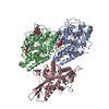


 Mass: 24.305 Da / Num. of mol.: 2 / Source method: obtained synthetically / Formula: Mg
 Mass: 523.180 Da / Num. of mol.: 1 / Source method: obtained synthetically / Formula: C10H16N5O14P3 / Comment: GTP, energy-carrying molecule*YM
 Type: RNA linking / Mass: 443.201 Da / Num. of mol.: 1 / Source method: obtained synthetically / Formula: C10H15N5O11P2 / Comment: GDP, energy-carrying molecule*YM
 Mass: 853.906 Da / Num. of mol.: 1 / Source method: obtained synthetically / Formula: C47H51NO14 / Comment: medication, chemotherapy*YM
 Mass: 427.201 Da / Num. of mol.: 1 / Source method: obtained synthetically / Formula: C10H15N5O10P2 / Comment: ADP, energy-carrying molecule*YM
 Sample preparation
 Electron microscopy imaging
 FIELD EMISSION GUN / Accelerating voltage: 200 kV / Illumination mode: FLOOD BEAM
 Processing