 Movie
Movie Controller

[English] 日本語
 Yorodumi
- PDB-2c9f: THE QUASI-ATOMIC MODEL OF THE ADENOVIRUS TYPE 3 PENTON DODECAHEDRON -
+ Open data

- Basic information
| Entry | Database: PDB / ID: 2c9f | ||||||
|---|---|---|---|---|---|---|---|
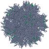
| Title | THE QUASI-ATOMIC MODEL OF THE ADENOVIRUS TYPE 3 PENTON DODECAHEDRON | ||||||
 Components Components |
| ||||||
 Keywords Keywords | VIRUS/VIRAL PROTEIN / VIRUS-VIRAL PROTEIN COMPLEX /  DODECAHEDRON / PENTON / FIBRE DODECAHEDRON / PENTON / FIBRE | ||||||
| Function / homology |  Function and homology information Function and homology informationT=25 icosahedral viral capsid / adhesion receptor-mediated virion attachment to host cell / viral capsid / clathrin-dependent endocytosis of virus by host cell / cell adhesion / symbiont entry into host cell / host cell nucleus / structural molecule activity / virion attachment to host cellSimilarity search - Function | ||||||
| Biological species |   HUMAN ADENOVIRUS TYPE 2 HUMAN ADENOVIRUS TYPE 2 | ||||||
| Method | ELECTRON MICROSCOPY / single particle reconstruction / cryo EM / Resolution: 16.5 Å | ||||||
 Authors Authors | Fuschiotti, P. / Schoehn, G. / Fender, P. / Fabry, C.M.S. / Hewat, E.A. / Chroboczek, J. / Ruigrok, R.W.H. / Conway, J.F. | ||||||
 Citation Citation | Journal: J Mol Biol / Year: 2006 Title: Structure of the dodecahedral penton particle from human adenovirus type 3. Authors: P Fuschiotti / G Schoehn / P Fender / C M S Fabry / E A Hewat / J Chroboczek / R W H Ruigrok / J F Conway /  Abstract: The sub-viral dodecahedral particle of human adenovirus type 3, composed of the viral penton base and fiber proteins, shares an important characteristic of the entire virus: it can attach to cells ...The sub-viral dodecahedral particle of human adenovirus type 3, composed of the viral penton base and fiber proteins, shares an important characteristic of the entire virus: it can attach to cells and penetrate them. Structure determination of the fiberless dodecahedron by cryo-electron microscopy to 9 Angstroms resolution reveals tightly bound pentamer subunits, with only minimal interfaces between penton bases stabilizing the fragile dodecahedron. The internal cavity of the dodecahedron is approximately 80 Angstroms in diameter, and the interior surface is accessible to solvent through perforations of approximately 20 Angstroms diameter between the pentamer towers. We observe weak density beneath pentamers that we attribute to a penton base peptide including residues 38-48. The intact amino-terminal domain appears to interfere with pentamer-pentamer interactions and its absence by mutation or proteolysis is essential for dodecamer assembly. Differences between the 9 Angstroms dodecahedron structure and the adenovirus serotype 2 (Ad2) crystallographic model correlate closely with differences in sequence. The 3D structure of the dodecahedron including fibers at 16 Angstroms resolution reveals extra density on the top of the penton base that can be attributed to the fiber N terminus. The fiber itself exhibits striations that correlate with features of the atomic structure of the partial Ad2 fiber and that represent a repeat motif present in the amino acid sequence. These new observations offer important insights into particle assembly and stability, as well as the practicality of using the dodecahedron in targeted drug delivery. The structural work provides a sound basis for manipulating the properties of this particle and thereby enhancing its value for such therapeutic use. | ||||||
| History |
|
- Structure visualization
| Movie |
Movie viewer |
|---|---|
| Structure viewer | Molecule: MolmilJmol/JSmol |
- Downloads & links
-Download
| PDBx/mmCIF format | 2c9f.cif.gz | 444.6 KB | Display | PDBx/mmCIF format |
|---|---|---|---|---|
| PDB format | pdb2c9f.ent.gz | 371 KB | Display | PDB format |
| PDBx/mmJSON format | 2c9f.json.gz | Tree view | PDBx/mmJSON format | |
| Others |  Other downloads Other downloads |
-Validation report
| Arichive directory | https://data.pdbj.org/pub/pdb/validation_reports/c9/2c9fftp://data.pdbj.org/pub/pdb/validation_reports/c9/2c9f | HTTPS FTP |
|---|
-Related structure data
| Related structure data |  1178MPC  1179FC  2c9gC C: citing same article ( M: map data used to model this data P: unfit; worng pairing?*YM F: fitted*YM |
|---|---|
| Similar structure data |


-Links
 PDBj
PDBj

- Assembly
| Deposited unit | 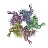
|
|---|---|
| 1 | x 12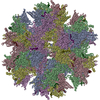
|
| 2 |
|
| 3 | 
|
| 4 | 
|
| 5 | 
|
| Symmetry | Point symmetry: (Schoenflies symbol: I (icosahedral)) |
-Components
| #1: Protein | / VIRION COMPONENT III / PENTON BASE PROTEIN / PIII Mass: 58218.684 Da / Num. of mol.: 5 / Fragment: RESIDUES 49-571 Source method: isolated from a genetically manipulated source Source: (gene. exp.) HUMAN ADENOVIRUS TYPE 2 / Production host:  TRICHOPLUSIA NI (cabbage looper) / References: UniProt: P03276 TRICHOPLUSIA NI (cabbage looper) / References: UniProt: P03276#2: Protein/peptide | / N-TERMINAL PEPTIDE OF THE FIBERMass: 2218.468 Da / Num. of mol.: 5 / Fragment: RESIDUES 1-19 / Source method: isolated from a natural source / Details: VIRION COMPONENT IV, FIBER PROTEIN / Source: (natural) HUMAN ADENOVIRUS TYPE 2 / References: UniProt: Q64831, UniProt: P03275*PLUS |
|---|
-Experimental details
-Experiment
| Experiment | Method: ELECTRON MICROSCOPY |
|---|---|
| EM experiment | Aggregation state: PARTICLE / 3D reconstruction method: single particle reconstruction |
- Sample preparation
| Component | Name: HUMAN ADENOVIRUS TYPE 3 ENTIRE DODECAHEDRAL PARTICLE / Type: VIRUS |
|---|---|
| Buffer solution | pH: 6.6 |
| Specimen | Conc.: 1 mg/ml / Embedding applied: NO / Shadowing applied: NO / Staining applied: NO / Vitrification applied: YES |
| Vitrification | Cryogen name: ETHANE / Details: LIQUID ETHANE |
- Electron microscopy imaging
| Microscopy | Model: FEI/PHILIPS CM200T |
|---|---|
| Electron gun | Electron source: LAB6 / Accelerating voltage: 200 kV / Illumination mode: FLOOD BEAM |
| Electron lens | Mode: BRIGHT FIELDBright-field microscopy / Nominal magnification: 38000 X / Calibrated magnification: 40930 X / Nominal defocus max: 2500 nm / Nominal defocus min: 1000 nm / Cs: 2 mm |
| Specimen holder | Temperature: 95 K / Tilt angle max: 0 ° / Tilt angle min: 0 ° |
| Image recording | Film or detector model: KODAK SO-163 FILM |
| Image scans | Num. digital images: 12 |
| Radiation wavelength | Relative weight: 1 |
- Processing
| EM software |
| ||||||||||||
|---|---|---|---|---|---|---|---|---|---|---|---|---|---|
| CTF correction | Details: AMPLITUDE, PHASE | ||||||||||||
| Symmetry | Point symmetry: I (icosahedral) | ||||||||||||
| 3D reconstruction | Method: POLAR FOURIER TRANSFROM, FOURIER BESSELS RECONSTRUCTION Resolution: 16.5 Å / Num. of particles: 1849 / Nominal pixel size: 1.84 Å / Actual pixel size: 1.71 Å Magnification calibration: COMPARISON WITH THE ATOMIC STRUCTURE Details: TO RECONSTITUTE THE ENTIRE DODECAHEDRON, YOU MUST APPLY THE 12 MATRICES TO THE ENTIRE PENTAMER. Symmetry type: POINT | ||||||||||||
| Atomic model building | Protocol: RIGID BODY FIT / Space: RECIPROCAL / Target criteria: RFACTOR, CORRELATION COEFFICIENT / Details: METHOD--RIGID BODY REFINEMENT PROTOCOL--X-RAY | ||||||||||||
| Refinement | Highest resolution: 16.5 Å | ||||||||||||
| Refinement step | Cycle: LAST / Highest resolution: 16.5 Å
|
