 Movie
Movie Controller


+ Open data

- Basic information
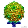
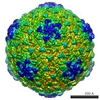
| Entry | Database: EMDB / ID: EMD-2965 | |||||||||
|---|---|---|---|---|---|---|---|---|---|---|
| Title | Cryo-EM structure of Ross River virus | |||||||||
 Map data Map data | Cryo-EM 3D reconstruction of Ross River virus | |||||||||
 Sample Sample |
| |||||||||
 Keywords Keywords | Ross River virus | |||||||||
| Biological species |  Ross River virus Ross River virus | |||||||||
| Method | single particle reconstruction / cryo EM / negative staining / Resolution: 15.2 Å | |||||||||
 Authors Authors | Chen C / Wang J C-Y / Rayaprolu V / Mukhopadhyay S / Zlotnick A | |||||||||
 Citation Citation | Journal: ACS Nano / Year: 2015 Title: Self-Assembly of an Alphavirus Core-like Particle Is Distinguished by Strong Intersubunit Association Energy and Structural Defects. Authors: Joseph Che-Yen Wang / Chao Chen / Vamseedhar Rayaprolu / Suchetana Mukhopadhyay / Adam Zlotnick /  Abstract: Weak association energy can lead to uniform nanostructures: defects can anneal due to subunit lability. What happens when strong association energy leads to particles where defects are trapped? ...Weak association energy can lead to uniform nanostructures: defects can anneal due to subunit lability. What happens when strong association energy leads to particles where defects are trapped? Alphaviruses are enveloped viruses whose icosahedral nucleocapsid core can assemble independently. We used a simplest case system to study Ross River virus (RRV) core-like particle (CLP) self-assembly using purified capsid protein and a short DNA oligomer. We find that capsid protein binds the oligomer with high affinity to form an assembly competent unit (U). Subsequently, U assembles with concentration dependence into CLPs. We determined that U-U pairwise interactions are very strong (ca. -6 kcal/mol) compared to other virus assembly systems. Assembled RRV CLPs appeared morphologically uniform and cryo-EM image reconstruction with imposed icosahedral symmetry yielded a T = 4 structure. However, 2D class averages of the CLPs show that virtually every class had disordered regions. These results suggested that irregular cores may be present in RRV virions. To test this hypothesis, we determined 2D class averages of RRV virions using authentic virions or only the core from intact virions isolated by computational masking. Virion-based class averages were symmetrical, geometric, and corresponded well to projections of image reconstructions. In core-based class averages, cores and envelope proteins in many classes were disordered. These results suggest that partly disordered components are common even in ostensibly well-ordered viruses, a biological realization of a patchy particle. Biological advantages of partly disordered complexes may arise from their ease of dissociation and asymmetry. | |||||||||
| History |
|
- Structure visualization
| Movie |
 Movie viewer Movie viewer |
|---|---|
| Structure viewer | EM map: SurfViewMolmilJmol/JSmol |
| Supplemental images |

- Downloads & links
-EMDB archive
| Map data | emd_2965.map.gz | 131.3 MB | EMDB map data format | |
|---|---|---|---|---|
| Header (meta data) | emd-2965-v30.xmlemd-2965.xml | 9.4 KB 9.4 KB | Display Display | EMDB header |
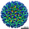
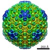
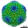
| Images | 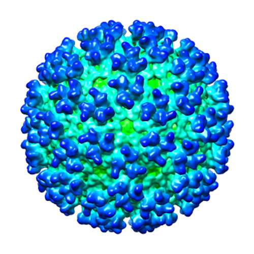 emd_2965.png emd_2965.png | 252.9 KB | ||
| Archive directory |  http://ftp.pdbj.org/pub/emdb/structures/EMD-2965ftp://ftp.pdbj.org/pub/emdb/structures/EMD-2965 http://ftp.pdbj.org/pub/emdb/structures/EMD-2965ftp://ftp.pdbj.org/pub/emdb/structures/EMD-2965 | HTTPS FTP |
-Validation report
| Summary document | emd_2965_validation.pdf.gz | 233.7 KB | Display | EMDB validaton report |
|---|---|---|---|---|
| Full document | emd_2965_full_validation.pdf.gz | 232.8 KB | Display | |
| Data in XML | emd_2965_validation.xml.gz | 7.7 KB | Display | |
| Arichive directory | https://ftp.pdbj.org/pub/emdb/validation_reports/EMD-2965ftp://ftp.pdbj.org/pub/emdb/validation_reports/EMD-2965 | HTTPS FTP |
-Related structure data


-Links
| EMDB pages | EMDB (EBI/PDBe) / EMDataResource |
|---|
-Map
| File | Download / File: emd_2965.map.gz / Format: CCP4 / Size: 319.5 MB / Type: IMAGE STORED AS FLOATING POINT NUMBER (4 BYTES) | ||||||||||||||||||||||||||||||||||||||||||||||||||||||||||||
|---|---|---|---|---|---|---|---|---|---|---|---|---|---|---|---|---|---|---|---|---|---|---|---|---|---|---|---|---|---|---|---|---|---|---|---|---|---|---|---|---|---|---|---|---|---|---|---|---|---|---|---|---|---|---|---|---|---|---|---|---|---|
| Annotation | Cryo-EM 3D reconstruction of Ross River virus | ||||||||||||||||||||||||||||||||||||||||||||||||||||||||||||
| Voxel size | X=Y=Z: 1.84 Å | ||||||||||||||||||||||||||||||||||||||||||||||||||||||||||||
| Density |
| ||||||||||||||||||||||||||||||||||||||||||||||||||||||||||||
| Symmetry | Space group: 1 | ||||||||||||||||||||||||||||||||||||||||||||||||||||||||||||
| Details | EMDB XML:
CCP4 map header:
| ||||||||||||||||||||||||||||||||||||||||||||||||||||||||||||
-Supplemental data
- Sample components
-Entire : Cryo-EM structure of Ross River virus
| Entire | Name: Cryo-EM structure of Ross River virus |
|---|---|
| Components |
|
-Supramolecule #1000: Cryo-EM structure of Ross River virus
| Supramolecule | Name: Cryo-EM structure of Ross River virus / type: sample / ID: 1000 Details: The virus was purified from Baby Hamster Kidney cells Number unique components: 1 |
|---|
-Supramolecule #1: Ross River virus
| Supramolecule | Name: Ross River virus / type: virus / ID: 1 / NCBI-ID: 11029 / Sci species name: Ross River virus / Virus type: VIRION / Virus isolate: OTHER / Virus enveloped: Yes / Virus empty: No |
|---|---|
| Host (natural) | Organism:  |
| Host system | Organism:  Mesocricetus auratus (golden hamster) / Recombinant cell: BHK-21 / Recombinant plasmid: cDNA clone of Ross River T48 virus Mesocricetus auratus (golden hamster) / Recombinant cell: BHK-21 / Recombinant plasmid: cDNA clone of Ross River T48 virus |
| Virus shell | Shell ID: 1 / T number (triangulation number): 4 |
-Experimental details
-Structure determination
| Method | negative staining, cryo EM |
|---|---|
 Processing Processing | single particle reconstruction |
| Aggregation state | particle |
-Sample preparation
| Buffer | pH: 7.4 / Details: 20 mM Hepes, 150 mM NaCl and 0.1 mM EDTA |
|---|---|
| Staining | Type: NEGATIVE / Details: cryo-EM |
| Grid | Details: 300 mesh copper grid with thin carbon support |
| Vitrification | Cryogen name: ETHANE / Chamber humidity: 100 % / Instrument: FEI VITROBOT MARK III / Method: wait 25 sec and blot for 4 sec before plunging |
- Electron microscopy
| Microscope | JEOL 3200FS |
|---|---|
| Temperature | Average: 100 K |
| Alignment procedure | Legacy - Astigmatism: Objective lens astigmatism was corrected at 60,000 times magnification |
| Specialist optics | Energy filter - Name: omega filter / Energy filter - Lower energy threshold: 0.0 eV / Energy filter - Upper energy threshold: 20.0 eV |
| Details | MDS |
| Date | Mar 15, 2015 |
| Image recording | Category: CCD / Film or detector model: GATAN ULTRASCAN 4000 (4k x 4k) / Number real images: 127 / Average electron dose: 25 e/Å2 |
| Electron beam | Acceleration voltage: 300 kV / Electron source:  FIELD EMISSION GUN FIELD EMISSION GUN |
| Electron optics | Illumination mode: FLOOD BEAM / Imaging mode: BRIGHT FIELD / Cs: 1.1 mm / Nominal defocus max: 4.01 µm / Nominal defocus min: 1.31 µm / Nominal magnification: 60000 |
| Sample stage | Specimen holder: 626 / Specimen holder model: GATAN LIQUID NITROGEN |
-Image processing
| Details | The particles were selected using e2boxer.py and the images were processed using auto3dem. The initial model was built de novo. |
|---|---|
| CTF correction | Details: per micrograph |
| Final reconstruction | Applied symmetry - Point group: I (icosahedral) / Algorithm: OTHER / Resolution.type: BY AUTHOR / Resolution: 15.2 Å / Resolution method: OTHER / Software - Name: auto3dem / Number images used: 6146 |
