cytochrome o ubiquinol oxidase complex / oxidoreduction-driven active transmembrane transporter activity / ubiquinol oxidase (H+-transporting) / cytochrome bo3 ubiquinol oxidase activity / aerobic electron transport chain / oxidoreductase activity, acting on diphenols and related substances as donors, oxygen as acceptor / ubiquinone binding / electron transport coupled proton transport / cytochrome-c oxidase activity / proton transmembrane transporter activity ...cytochrome o ubiquinol oxidase complex / oxidoreduction-driven active transmembrane transporter activity / ubiquinol oxidase (H+-transporting) / cytochrome bo3 ubiquinol oxidase activity / aerobic electron transport chain / oxidoreductase activity, acting on diphenols and related substances as donors, oxygen as acceptor / ubiquinone binding / electron transport coupled proton transport / cytochrome-c oxidase activity / proton transmembrane transporter activity / ATP synthesis coupled electron transport / respirasome / aerobic respiration / respiratory electron transport chain / electron transfer activity / copper ion binding / heme binding / plasma membrane Similarity search - Function
Cytochrome o ubiquinol oxidase, subunit III / Cytochrome o ubiquinol oxidase subunit IV / Cytochrome o ubiquinol oxidase, subunit I / Ubiquinol oxidase subunit III domain / Cytochrome C oxidase subunit IV, prokaryotes / COX aromatic rich motif / Prokaryotic Cytochrome C oxidase subunit IV / COX Aromatic Rich Motif / Cytochrome o ubiquinol oxidase subunit II / Ubiquinol oxidase subunit 2, cupredoxin domain ...Cytochrome o ubiquinol oxidase, subunit III / Cytochrome o ubiquinol oxidase subunit IV / Cytochrome o ubiquinol oxidase, subunit I / Ubiquinol oxidase subunit III domain / Cytochrome C oxidase subunit IV, prokaryotes / COX aromatic rich motif / Prokaryotic Cytochrome C oxidase subunit IV / COX Aromatic Rich Motif / Cytochrome o ubiquinol oxidase subunit II / Ubiquinol oxidase subunit 2, cupredoxin domain / Cytochrome c oxidase subunit III / Cytochrome c oxidase subunit III-like / Cytochrome c oxidase, subunit III, 4-helical bundle / Cytochrome c oxidase subunit III / Heme-copper oxidase subunit III family profile. / Cytochrome c oxidase subunit III-like superfamily / Cytochrome C oxidase subunit II, transmembrane domain / Cytochrome oxidase subunit II transmembrane region profile. / Cytochrome c/quinol oxidase subunit II / Cytochrome C oxidase subunit II, transmembrane domain superfamily / Cytochrome c oxidase, subunit I, copper-binding site / Heme-copper oxidase catalytic subunit, copper B binding region signature. / Cytochrome c oxidase-like, subunit I domain / Cytochrome oxidase subunit I profile. / Cytochrome c oxidase subunit I / Cytochrome c oxidase-like, subunit I superfamily / Cytochrome C and Quinol oxidase polypeptide I / Cytochrome C oxidase subunit II, periplasmic domain / Cytochrome c oxidase subunit II-like C-terminal / Cytochrome oxidase subunit II copper A binding domain profile. / Cupredoxin / Prokaryotic membrane lipoprotein lipid attachment site profile. Similarity search - Domain/homology
National Institutes of Health/National Institute Of Allergy and Infectious Diseases (NIH/NIAID)
United States
Citation
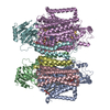
Journal: Protein Sci / Year: 2023 Title: Monomer and dimer structures of cytochrome bo ubiquinol oxidase from Escherichia coli. Authors: Yirui Guo / Elina Karimullina / Tabitha Emde / Zbyszek Otwinowski / Dominika Borek / Alexei Savchenko / Abstract: The Escherichia coli cytochrome bo ubiquinol oxidase is a four-subunit heme-copper oxidase that serves as a proton pump in the E. coli aerobic respiratory chain. Despite many mechanistic studies, it ...The Escherichia coli cytochrome bo ubiquinol oxidase is a four-subunit heme-copper oxidase that serves as a proton pump in the E. coli aerobic respiratory chain. Despite many mechanistic studies, it is unclear whether this ubiquinol oxidase functions as a monomer, or as a dimer in a manner similar to its eukaryotic counterparts-the mitochondrial electron transport complexes. In this study, we determined the monomeric and dimeric structures of the E. coli cytochrome bo ubiquinol oxidase reconstituted in amphipol by cryogenic electron microscopy single particle reconstruction (cryo-EM SPR) to a resolution of 3.15 and 3.46 Å, respectively. We have discovered that the protein can form a dimer with C2 symmetry, with the dimerization interface maintained by interactions between the subunit II of one monomer and the subunit IV of the other monomer. Moreover, the dimerization does not induce significant structural changes in the monomers, except the movement of a loop in subunit IV (residues 67-74).
In the structure databanks used in Yorodumi, some data are registered as the other names, "COVID-19 virus" and "2019-nCoV". Here are the details of the virus and the list of structure data.
Jan 31, 2019. EMDB accession codes are about to change! (news from PDBe EMDB page)
EMDB accession codes are about to change! (news from PDBe EMDB page)
The allocation of 4 digits for EMDB accession codes will soon come to an end. Whilst these codes will remain in use, new EMDB accession codes will include an additional digit and will expand incrementally as the available range of codes is exhausted. The current 4-digit format prefixed with “EMD-” (i.e. EMD-XXXX) will advance to a 5-digit format (i.e. EMD-XXXXX), and so on. It is currently estimated that the 4-digit codes will be depleted around Spring 2019, at which point the 5-digit format will come into force.
The EM Navigator/Yorodumi systems omit the EMD- prefix.
Related info.:Q: What is EMD? / ID/Accession-code notation in Yorodumi/EM Navigator
Yorodumi is a browser for structure data from EMDB, PDB, SASBDB, etc.
This page is also the successor to EM Navigator detail page, and also detail information page/front-end page for Omokage search.
The word "yorodu" (or yorozu) is an old Japanese word meaning "ten thousand". "mi" (miru) is to see.
Related info.:EMDB / PDB / SASBDB / Comparison of 3 databanks / Yorodumi Search / Aug 31, 2016. New EM Navigator & Yorodumi / Yorodumi Papers / Jmol/JSmol / Function and homology information / Changes in new EM Navigator and Yorodumi
 Movie
Movie Controller


 Open data
 Basic information
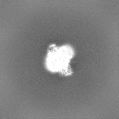
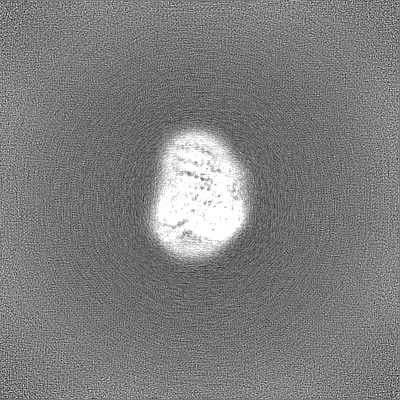
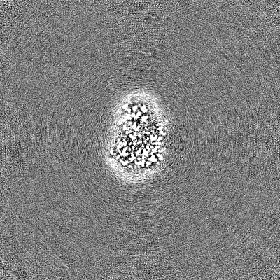
 Map data
 Sample
 Function and homology information
 ubiquinol oxidase (H+-transporting) / cytochrome bo3 ubiquinol oxidase activity / aerobic electron transport chain / oxidoreductase activity, acting on diphenols and related substances as donors, oxygen as acceptor /
 Authors
 United States, 1 items
  Citation
 Structure visualization
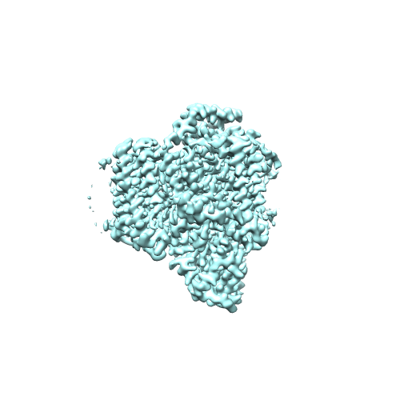
 Downloads & links
 emd_28877.png
 http://ftp.pdbj.org/pub/emdb/structures/EMD-28877
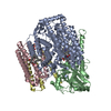


 Z
Z Y
Y X
X


 Sample components


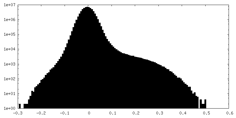
 Processing
 Electron microscopy
