 Movie
Movie Controller

[English] 日本語
 Yorodumi
- EMDB-28724: Cryo-EM structure of 4 insulins bound full-length mouse IR mutant... -
+ Open data

- Basic information
| Entry |  | |||||||||
|---|---|---|---|---|---|---|---|---|---|---|
| Title | Cryo-EM structure of 4 insulins bound full-length mouse IR mutant with physically decoupled alpha CTs (C684S/C685S/C687S, denoted as IR-3CS) Asymmetric conformation 2 | |||||||||
 Map data Map data | Cryo-EM structure of 4 insulins bound full-length mouse IR mutant with physically decoupled alpha CTs (C684S/C685S/C687S; denoted as IR-3CS) Asymmetric conformation 2 | |||||||||
 Sample Sample |
| |||||||||
| Function / homology |  Function and homology information Function and homology informationSignaling by Insulin receptor / Insulin receptor recycling /  yolk / IRS activation / Insulin receptor signalling cascade / Signal attenuation / 3-phosphoinositide-dependent protein kinase binding / positive regulation of glycoprotein biosynthetic process / lipoic acid binding / regulation of hydrogen peroxide metabolic process ...Signaling by Insulin receptor / Insulin receptor recycling / yolk / IRS activation / Insulin receptor signalling cascade / Signal attenuation / 3-phosphoinositide-dependent protein kinase binding / positive regulation of glycoprotein biosynthetic process / lipoic acid binding / regulation of hydrogen peroxide metabolic process / regulation of female gonad development / positive regulation of meiotic cell cycle / PI5P, PP2A and IER3 Regulate PI3K/AKT Signaling / positive regulation of developmental growth / insulin-like growth factor II binding / male sex determination / exocrine pancreas development / insulin receptor complex / insulin-like growth factor I binding / insulin receptor activity / nuclear lumen / positive regulation of protein-containing complex disassembly / cargo receptor activity / dendritic spine maintenance / PTB domain binding / insulin binding / negative regulation of NAD(P)H oxidase activity / neuronal cell body membrane / adrenal gland development / negative regulation of glycogen catabolic process / regulation of cellular amino acid metabolic process / Signaling by Insulin receptor / IRS activation / nitric oxide-cGMP-mediated signaling / Insulin processing / negative regulation of fatty acid metabolic process / negative regulation of feeding behavior / regulation of protein secretion / amyloid-beta clearance / positive regulation of peptide hormone secretion / positive regulation of respiratory burst / Regulation of gene expression in beta cells / regulation of embryonic development / positive regulation of receptor internalization / negative regulation of acute inflammatory response / alpha-beta T cell activation / negative regulation of respiratory burst involved in inflammatory response / insulin receptor substrate binding / positive regulation of dendritic spine maintenance / Synthesis, secretion, and deacylation of Ghrelin / epidermis development / positive regulation of glycogen biosynthetic process / negative regulation of protein secretion / Signal attenuation / FOXO-mediated transcription of oxidative stress, metabolic and neuronal genes / response to tumor necrosis factor / negative regulation of gluconeogenesis / positive regulation of nitric oxide mediated signal transduction / fatty acid homeostasis / regulation of protein localization to plasma membrane / phosphatidylinositol 3-kinase binding / COPI-mediated anterograde transport / negative regulation of lipid catabolic process / positive regulation of lipid biosynthetic process / negative regulation of oxidative stress-induced intrinsic apoptotic signaling pathway / heart morphogenesis / positive regulation of insulin receptor signaling pathway / positive regulation of phosphorylation / negative regulation of reactive oxygen species biosynthetic process / transport vesicle / positive regulation of protein autophosphorylation / dendrite membrane / Insulin receptor recycling / insulin-like growth factor receptor binding / NPAS4 regulates expression of target genes / positive regulation of protein metabolic process / neuron projection maintenance / positive regulation of brown fat cell differentiation / endoplasmic reticulum-Golgi intermediate compartment membrane / activation of protein kinase B activity / positive regulation of glycolytic process / response to nutrient levels / Insulin receptor signalling cascade / receptor-mediated endocytosis / positive regulation of mitotic nuclear division / negative regulation of protein phosphorylation / Regulation of insulin secretion / positive regulation of long-term synaptic potentiation / caveola / endosome lumen / positive regulation of cytokine production / acute-phase response / positive regulation of protein secretion / positive regulation of nitric-oxide synthase activity / regulation of transmembrane transporter activity / positive regulation of cell differentiation / positive regulation of glucose import / negative regulation of proteolysis / animal organ morphogenesis / regulation of synaptic plasticity yolk / IRS activation / Insulin receptor signalling cascade / Signal attenuation / 3-phosphoinositide-dependent protein kinase binding / positive regulation of glycoprotein biosynthetic process / lipoic acid binding / regulation of hydrogen peroxide metabolic process ...Signaling by Insulin receptor / Insulin receptor recycling / yolk / IRS activation / Insulin receptor signalling cascade / Signal attenuation / 3-phosphoinositide-dependent protein kinase binding / positive regulation of glycoprotein biosynthetic process / lipoic acid binding / regulation of hydrogen peroxide metabolic process / regulation of female gonad development / positive regulation of meiotic cell cycle / PI5P, PP2A and IER3 Regulate PI3K/AKT Signaling / positive regulation of developmental growth / insulin-like growth factor II binding / male sex determination / exocrine pancreas development / insulin receptor complex / insulin-like growth factor I binding / insulin receptor activity / nuclear lumen / positive regulation of protein-containing complex disassembly / cargo receptor activity / dendritic spine maintenance / PTB domain binding / insulin binding / negative regulation of NAD(P)H oxidase activity / neuronal cell body membrane / adrenal gland development / negative regulation of glycogen catabolic process / regulation of cellular amino acid metabolic process / Signaling by Insulin receptor / IRS activation / nitric oxide-cGMP-mediated signaling / Insulin processing / negative regulation of fatty acid metabolic process / negative regulation of feeding behavior / regulation of protein secretion / amyloid-beta clearance / positive regulation of peptide hormone secretion / positive regulation of respiratory burst / Regulation of gene expression in beta cells / regulation of embryonic development / positive regulation of receptor internalization / negative regulation of acute inflammatory response / alpha-beta T cell activation / negative regulation of respiratory burst involved in inflammatory response / insulin receptor substrate binding / positive regulation of dendritic spine maintenance / Synthesis, secretion, and deacylation of Ghrelin / epidermis development / positive regulation of glycogen biosynthetic process / negative regulation of protein secretion / Signal attenuation / FOXO-mediated transcription of oxidative stress, metabolic and neuronal genes / response to tumor necrosis factor / negative regulation of gluconeogenesis / positive regulation of nitric oxide mediated signal transduction / fatty acid homeostasis / regulation of protein localization to plasma membrane / phosphatidylinositol 3-kinase binding / COPI-mediated anterograde transport / negative regulation of lipid catabolic process / positive regulation of lipid biosynthetic process / negative regulation of oxidative stress-induced intrinsic apoptotic signaling pathway / heart morphogenesis / positive regulation of insulin receptor signaling pathway / positive regulation of phosphorylation / negative regulation of reactive oxygen species biosynthetic process / transport vesicle / positive regulation of protein autophosphorylation / dendrite membrane / Insulin receptor recycling / insulin-like growth factor receptor binding / NPAS4 regulates expression of target genes / positive regulation of protein metabolic process / neuron projection maintenance / positive regulation of brown fat cell differentiation / endoplasmic reticulum-Golgi intermediate compartment membrane / activation of protein kinase B activity / positive regulation of glycolytic process / response to nutrient levels / Insulin receptor signalling cascade / receptor-mediated endocytosis / positive regulation of mitotic nuclear division / negative regulation of protein phosphorylation / Regulation of insulin secretion / positive regulation of long-term synaptic potentiation / caveola / endosome lumen / positive regulation of cytokine production / acute-phase response / positive regulation of protein secretion / positive regulation of nitric-oxide synthase activity / regulation of transmembrane transporter activity / positive regulation of cell differentiation / positive regulation of glucose import / negative regulation of proteolysis / animal organ morphogenesis / regulation of synaptic plasticitySimilarity search - Function | |||||||||
| Biological species |  Mus musculus (house mouse) / Mus musculus (house mouse) /  Homo sapiens (human) Homo sapiens (human) | |||||||||
| Method | single particle reconstruction / cryo EM / Resolution: 4.9 Å | |||||||||
 Authors Authors | Li J / Wu JY / Hall C / Bai XC / Choi E | |||||||||
| Funding support |  United States, 2 items United States, 2 items
| |||||||||
 Citation Citation | Journal: Elife / Year: 2022 Title: Molecular basis for the role of disulfide-linked αCTs in the activation of insulin-like growth factor 1 receptor and insulin receptor. Authors: Jie Li / Jiayi Wu / Catherine Hall / Xiao-Chen Bai / Eunhee Choi / Abstract: The insulin receptor (IR) and insulin-like growth factor 1 receptor (IGF1R) control metabolic homeostasis and cell growth and proliferation. The IR and IGF1R form similar disulfide bonds linked ...The insulin receptor (IR) and insulin-like growth factor 1 receptor (IGF1R) control metabolic homeostasis and cell growth and proliferation. The IR and IGF1R form similar disulfide bonds linked homodimers in the apo-state; however, their ligand binding properties and the structures in the active state differ substantially. It has been proposed that the disulfide-linked C-terminal segment of α-chain (αCTs) of the IR and IGF1R control the cooperativity of ligand binding and regulate the receptor activation. Nevertheless, the molecular basis for the roles of disulfide-linked αCTs in IR and IGF1R activation are still unclear. Here, we report the cryo-EM structures of full-length mouse IGF1R/IGF1 and IR/insulin complexes with modified αCTs that have increased flexibility. Unlike the -shaped asymmetric IGF1R dimer with a single IGF1 bound, the IGF1R with the enhanced flexibility of αCTs can form a -shaped symmetric dimer with two IGF1s bound. Meanwhile, the IR with non-covalently linked αCTs predominantly adopts an asymmetric conformation with four insulins bound, which is distinct from the -shaped symmetric IR. Using cell-based experiments, we further showed that both IGF1R and IR with the modified αCTs cannot activate the downstream signaling potently. Collectively, our studies demonstrate that the certain structural rigidity of disulfide-linked αCTs is critical for optimal IR and IGF1R signaling activation. | |||||||||
| History |
|
- Structure visualization
| Supplemental images |
|---|

- Downloads & links
-EMDB archive
| Map data | emd_28724.map.gz | 61.8 MB | EMDB map data format | |
|---|---|---|---|---|
| Header (meta data) | emd-28724-v30.xmlemd-28724.xml | 17.7 KB 17.7 KB | Display Display | EMDB header |
| Images | 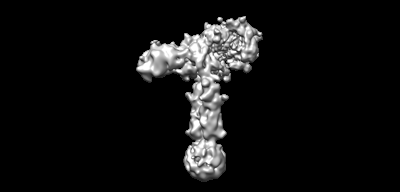 emd_28724.png emd_28724.png | 20.5 KB | ||
| Others | emd_28724_half_map_1.map.gzemd_28724_half_map_2.map.gz | 80.7 MB 80.7 MB | ||
| Archive directory |  http://ftp.pdbj.org/pub/emdb/structures/EMD-28724ftp://ftp.pdbj.org/pub/emdb/structures/EMD-28724 http://ftp.pdbj.org/pub/emdb/structures/EMD-28724ftp://ftp.pdbj.org/pub/emdb/structures/EMD-28724 | HTTPS FTP |
-Related structure data
| Related structure data |  8eyyMC  8eyrC  8eyxC  8ez0C C: citing same article ( M: atomic model generated by this map |
|---|---|
| Similar structure data |


-Links
| EMDB pages | EMDB (EBI/PDBe) / EMDataResource |
|---|---|
| Related items in Molecule of the Month |


-Map
| File | Download / File: emd_28724.map.gz / Format: CCP4 / Size: 103 MB / Type: IMAGE STORED AS FLOATING POINT NUMBER (4 BYTES) | ||||||||||||||||||||
|---|---|---|---|---|---|---|---|---|---|---|---|---|---|---|---|---|---|---|---|---|---|
| Annotation | Cryo-EM structure of 4 insulins bound full-length mouse IR mutant with physically decoupled alpha CTs (C684S/C685S/C687S; denoted as IR-3CS) Asymmetric conformation 2 | ||||||||||||||||||||
| Voxel size | X=Y=Z: 1.08 Å | ||||||||||||||||||||
| Density |
| ||||||||||||||||||||
| Symmetry | Space group: 1 | ||||||||||||||||||||
| Details | EMDB XML:
|
-Supplemental data
-Half map: Cryo-EM structure of 4 insulins bound full-length mouse...
| File | emd_28724_half_map_1.map | ||||||||||||
|---|---|---|---|---|---|---|---|---|---|---|---|---|---|
| Annotation | Cryo-EM structure of 4 insulins bound full-length mouse IR mutant with physically decoupled alpha CTs (C684S/C685S/C687S; denoted as IR-3CS) Asymmetric conformation 2 | ||||||||||||
| Projections & Slices |
| ||||||||||||
| Density Histograms |
 Z
Z Y
Y X
X


-Half map: Cryo-EM structure of 4 insulins bound full-length mouse...
| File | emd_28724_half_map_2.map | ||||||||||||
|---|---|---|---|---|---|---|---|---|---|---|---|---|---|
| Annotation | Cryo-EM structure of 4 insulins bound full-length mouse IR mutant with physically decoupled alpha CTs (C684S/C685S/C687S; denoted as IR-3CS) Asymmetric conformation 2 | ||||||||||||
| Projections & Slices |
| ||||||||||||
| Density Histograms |


- Sample components
-Entire : Cryo-EM structure of 4 insulins bound full-length mouse IR mutant...
| Entire | Name: Cryo-EM structure of 4 insulins bound full-length mouse IR mutant with physically decoupled alpha CTs (C684S/C685S/C687S; denoted as IR-3CS) Asymmetric conformation 2 |
|---|---|
| Components |
|
-Supramolecule #1: Cryo-EM structure of 4 insulins bound full-length mouse IR mutant...
| Supramolecule | Name: Cryo-EM structure of 4 insulins bound full-length mouse IR mutant with physically decoupled alpha CTs (C684S/C685S/C687S; denoted as IR-3CS) Asymmetric conformation 2 type: complex / Chimera: Yes / ID: 1 / Parent: 0 / Macromolecule list: all |
|---|---|
| Source (natural) | Organism: Mus musculus (house mouse) |
-Macromolecule #1: Insulin receptor
| Macromolecule | Name: Insulin receptor / type: protein_or_peptide / ID: 1 / Number of copies: 2 / Enantiomer: LEVO / EC number: receptor protein-tyrosine kinase |
|---|---|
| Source (natural) | Organism: Mus musculus (house mouse) |
| Molecular weight | Theoretical: 153.184406 KDa |
| Recombinant expression | Organism: Homo sapiens (human) |
| Sequence | String: HLYPGEVCPG MDIRNNLTRL HELENCSVIE GHLQILLMFK TRPEDFRDLS FPKLIMITDY LLLFRVYGLE SLKDLFPNLT VIRGSRLFF NYALVIFEMV HLKELGLYNL MNITRGSVRI EKNNELCYLA TIDWSRILDS VEDNYIVLNK DDNEECGDVC P GTAKGKTN ...String: HLYPGEVCPG MDIRNNLTRL HELENCSVIE GHLQILLMFK TRPEDFRDLS FPKLIMITDY LLLFRVYGLE SLKDLFPNLT VIRGSRLFF NYALVIFEMV HLKELGLYNL MNITRGSVRI EKNNELCYLA TIDWSRILDS VEDNYIVLNK DDNEECGDVC P GTAKGKTN CPATVINGQF VERCWTHSHC QKVCPTICKS HGCTAEGLCC HKECLGNCSE PDDPTKCVAC RNFYLDGQCV ET CPPPYYH FQDWRCVNFS FCQDLHFKCR NSRKPGCHQY VIHNNKCIPE CPSGYTMNSS NLMCTPCLGP CPKVCQILEG EKT IDSVTS AQELRGCTVI NGSLIINIRG GNNLAAELEA NLGLIEEISG FLKIRRSYAL VSLSFFRKLH LIRGETLEIG NYSF YALDN QNLRQLWDWS KHNLTITQGK LFFHYNPKLC LSEIHKMEEV SGTKGRQERN DIALKTNGDQ ASCENELLKF SFIRT SFDK ILLRWEPYWP PDFRDLLGFM LFYKEAPYQN VTEFDGQDAC GSNSWTVVDI DPPQRSNDPK SQTPSHPGWL MRGLKP WTQ YAIFVKTLVT FSDERRTYGA KSDIIYVQTD ATNPSVPLDP ISVSNSSSQI ILKWKPPSDP NGNITHYLVY WERQAED SE LFELDYCLKG LKLPSRTWSP PFESDDSQKH NQSEYDDSAS ESSSSPKTDS QILKELEESS FRKTFEDYLH NVVFVPRP S RKRRSLEEVG NVTATTLTLP DFPNVSSTIV PTSQEEHRPF EKVVNKESLV ISGLRHFTGY RIELQACNQD SPDERCSVA AYVSARTMPE AKADDIVGPV THEIFENNVV HLMWQEPKEP NGLIVLYEVS YRRYGDEELH LCVSRKHFAL ERGCRLRGLS PGNYSVRVR ATSLAGNGSW TEPTYFYVTD YLDVPSNIAK IIIGPLIFVF LFSVVIGSIY LFLRKRQPDG PMGPLYASSN P EYLSASDV FPSSVYVPDE WEVPREKITL LRELGQGSFG MVYEGNAKDI IKGEAETRVA VKTVNESASL RERIEFLNEA SV MKGFTCH HVVRLLGVVS KGQPTLVVME LMAHGDLKSH LRSLRPDAEN NPGRPPPTLQ EMIQMTAEIA DGMAYLNAKK FVH RDLAAR NCMVAHDFTV KIGDFGMTRD IYETDYYRKG GKGLLPVRWM SPESLKDGVF TASSDMWSFG VVLWEITSLA EQPY QGLSN EQVLKFVMDG GYLDPPDNCP ERLTDLMRMC WQFNPKMRPT FLEIVNLLKD DLHPSFPEVS FFYSEENKAP ESEEL EMEF EDMENVPLDR SSHCQREEAG GREGGSSLSI KRTYDEHIPY THMNGGKKNG RVLTLPRSNP S |
-Macromolecule #2: Insulin
| Macromolecule | Name: Insulin / type: protein_or_peptide / ID: 2 / Number of copies: 4 / Enantiomer: LEVO |
|---|---|
| Source (natural) | Organism: Homo sapiens (human) |
| Molecular weight | Theoretical: 11.989862 KDa |
| Recombinant expression | Organism: Saccharomyces cerevisiae |
| Sequence | String: MALWMRLLPL LALLALWGPD PAAAFVNQHL CGSHLVEALY LVCGERGFFY TPKTRREAED LQVGQVELGG GPGAGSLQPL ALEGSLQKR GIVEQCCTSI CSLYQLENYC N |
-Experimental details
-Structure determination
| Method | cryo EM |
|---|---|
 Processing Processing | single particle reconstruction |
| Aggregation state | particle |
-Sample preparation
| Concentration | 6 mg/mL |
|---|---|
| Buffer | pH: 7.4 |
| Vitrification | Cryogen name: ETHANE |
- Electron microscopy
| Microscope | FEI TITAN KRIOS |
|---|---|
| Electron beam | Acceleration voltage: 300 kV / Electron source: FIELD EMISSION GUN |
| Electron optics | Illumination mode: FLOOD BEAM / Imaging mode: BRIGHT FIELDBright-field microscopy / Nominal defocus max: 2.6 µm / Nominal defocus min: 1.6 µm |
| Image recording | Film or detector model: GATAN K3 BIOQUANTUM (6k x 4k) / Average electron dose: 60.0 e/Å2 |
| Experimental equipment |  Model: Titan Krios / Image courtesy: FEI Company |
-Image processing
| Particle selection | Number selected: 3283617 |
|---|---|
| Initial angle assignment | Type: PROJECTION MATCHING |
| Final angle assignment | Type: PROJECTION MATCHING |
| Final reconstruction | Applied symmetry - Point group: C1 (asymmetric) / Resolution.type: BY AUTHOR / Resolution: 4.9 Å / Resolution method: FSC 0.143 CUT-OFF / Software - Name: RELION / Number images used: 104347 |
