 Movie
Movie Controller


+ Open data

- Basic information
| Entry | Database: EMDB / ID: EMD-2855 | |||||||||
|---|---|---|---|---|---|---|---|---|---|---|
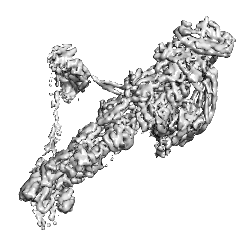
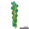
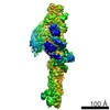
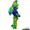
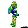
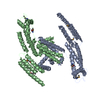
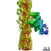
| Title | CryoEM structure of dynactin complex with p150-glued projection | |||||||||
 Map data Map data | CryoEM reconstruction of dynactin complex with clear extra density of p150-glued projection at 8.6 angstrom resolution. | |||||||||
 Sample Sample |
| |||||||||
 Keywords Keywords |  dynactin / dynein co-factor / actin-like filament / cellular cargo transport dynactin / dynein co-factor / actin-like filament / cellular cargo transport | |||||||||
| Function / homology |  Function and homology informationRegulation of PLK1 Activity at G2/M Transition / Loss of Nlp from mitotic centrosomes / Recruitment of mitotic centrosome proteins and complexes / Loss of proteins required for interphase microtubule organization from the centrosome / Anchoring of the basal body to the plasma membrane / AURKA Activation by TPX2 / positive regulation of neuromuscular junction development / centriolar subdistal appendage / centriole-centriole cohesion / ventral spinal cord development ...Regulation of PLK1 Activity at G2/M Transition / Loss of Nlp from mitotic centrosomes / Recruitment of mitotic centrosome proteins and complexes / Loss of proteins required for interphase microtubule organization from the centrosome / Anchoring of the basal body to the plasma membrane / AURKA Activation by TPX2 / positive regulation of neuromuscular junction development / centriolar subdistal appendage / centriole-centriole cohesion / ventral spinal cord development / microtubule anchoring at centrosome / melanosome transport / retromer complex / nuclear membrane disassembly / microtubule plus-end / positive regulation of microtubule nucleation / dynein complex / non-motile cilium assembly / Recruitment of NuMA to mitotic centrosomes / HSP90 chaperone cycle for steroid hormone receptors (SHR) in the presence of ligand / COPI-independent Golgi-to-ER retrograde traffic / MHC class II antigen presentation / retrograde transport, endosome to Golgi / nuclear migration / COPI-mediated anterograde transport / microtubule associated complex / motor behavior / neuromuscular process / neuromuscular junction development / intercellular bridge / cell leading edge / establishment of mitotic spindle orientation / regulation of mitotic spindle organization / neuron projection maintenance / centriole / ciliary basal body / mitotic spindle / kinetochore / spindle pole / neuron cellular homeostasis / nuclear envelope / cell cortex / microtubule binding / cell division / axon / centrosome / neuronal cell body / protein kinase binding / cytosol Function and homology informationRegulation of PLK1 Activity at G2/M Transition / Loss of Nlp from mitotic centrosomes / Recruitment of mitotic centrosome proteins and complexes / Loss of proteins required for interphase microtubule organization from the centrosome / Anchoring of the basal body to the plasma membrane / AURKA Activation by TPX2 / positive regulation of neuromuscular junction development / centriolar subdistal appendage / centriole-centriole cohesion / ventral spinal cord development ...Regulation of PLK1 Activity at G2/M Transition / Loss of Nlp from mitotic centrosomes / Recruitment of mitotic centrosome proteins and complexes / Loss of proteins required for interphase microtubule organization from the centrosome / Anchoring of the basal body to the plasma membrane / AURKA Activation by TPX2 / positive regulation of neuromuscular junction development / centriolar subdistal appendage / centriole-centriole cohesion / ventral spinal cord development / microtubule anchoring at centrosome / melanosome transport / retromer complex / nuclear membrane disassembly / microtubule plus-end / positive regulation of microtubule nucleation / dynein complex / non-motile cilium assembly / Recruitment of NuMA to mitotic centrosomes / HSP90 chaperone cycle for steroid hormone receptors (SHR) in the presence of ligand / COPI-independent Golgi-to-ER retrograde traffic / MHC class II antigen presentation / retrograde transport, endosome to Golgi / nuclear migration / COPI-mediated anterograde transport / microtubule associated complex / motor behavior / neuromuscular process / neuromuscular junction development / intercellular bridge / cell leading edge / establishment of mitotic spindle orientation / regulation of mitotic spindle organization / neuron projection maintenance / centriole / ciliary basal body / mitotic spindle / kinetochore / spindle pole / neuron cellular homeostasis / nuclear envelope / cell cortex / microtubule binding / cell division / axon / centrosome / neuronal cell body / protein kinase binding / cytosolSimilarity search - Function | |||||||||
| Biological species |  Sus scrofa domesticus (domestic pig) Sus scrofa domesticus (domestic pig) | |||||||||
| Method | single particle reconstruction / cryo EM / Resolution: 8.6 Å | |||||||||
 Authors Authors | Zhang K / Urnavicius L / Diamant AG / Motz C / Schlager MA / Yu M / Patel NA / Robinson CV / Carter AP | |||||||||
 Citation Citation | Journal: Science / Year: 2015 Title: The structure of the dynactin complex and its interaction with dynein. Authors: Linas Urnavicius / Kai Zhang / Aristides G Diamant / Carina Motz / Max A Schlager / Minmin Yu / Nisha A Patel / Carol V Robinson / Andrew P Carter /  Abstract: Dynactin is an essential cofactor for the microtubule motor cytoplasmic dynein-1. We report the structure of the 23-subunit dynactin complex by cryo-electron microscopy to 4.0 angstroms. Our ...Dynactin is an essential cofactor for the microtubule motor cytoplasmic dynein-1. We report the structure of the 23-subunit dynactin complex by cryo-electron microscopy to 4.0 angstroms. Our reconstruction reveals how dynactin is built around a filament containing eight copies of the actin-related protein Arp1 and one of β-actin. The filament is capped at each end by distinct protein complexes, and its length is defined by elongated peptides that emerge from the α-helical shoulder domain. A further 8.2 angstrom structure of the complex between dynein, dynactin, and the motility-inducing cargo adaptor Bicaudal-D2 shows how the translational symmetry of the dynein tail matches that of the dynactin filament. The Bicaudal-D2 coiled coil runs between dynein and dynactin to stabilize the mutually dependent interactions between all three components. | |||||||||
| History |
|
- Structure visualization
| Movie |
Movie viewer |
|---|---|
| Structure viewer | EM map: SurfViewMolmilJmol/JSmol |
| Supplemental images |
- Downloads & links
-EMDB archive
| Map data | emd_2855.map.gz | 14.4 MB | EMDB map data format | |
|---|---|---|---|---|
| Header (meta data) | emd-2855-v30.xmlemd-2855.xml | 18.2 KB 18.2 KB | Display Display | EMDB header |
| Images |  image2855.png image2855.png | 134.5 KB | ||
| Archive directory |  http://ftp.pdbj.org/pub/emdb/structures/EMD-2855ftp://ftp.pdbj.org/pub/emdb/structures/EMD-2855 http://ftp.pdbj.org/pub/emdb/structures/EMD-2855ftp://ftp.pdbj.org/pub/emdb/structures/EMD-2855 | HTTPS FTP |
-Related structure data
-Links
| EMDB pages | EMDB (EBI/PDBe) / EMDataResource |
|---|
-Map
| File | Download / File: emd_2855.map.gz / Format: CCP4 / Size: 173.8 MB / Type: IMAGE STORED AS FLOATING POINT NUMBER (4 BYTES) | ||||||||||||||||||||||||||||||||||||||||||||||||||||||||||||
|---|---|---|---|---|---|---|---|---|---|---|---|---|---|---|---|---|---|---|---|---|---|---|---|---|---|---|---|---|---|---|---|---|---|---|---|---|---|---|---|---|---|---|---|---|---|---|---|---|---|---|---|---|---|---|---|---|---|---|---|---|---|
| Annotation | CryoEM reconstruction of dynactin complex with clear extra density of p150-glued projection at 8.6 angstrom resolution. | ||||||||||||||||||||||||||||||||||||||||||||||||||||||||||||
| Voxel size | X=Y=Z: 1.7 Å | ||||||||||||||||||||||||||||||||||||||||||||||||||||||||||||
| Density |
| ||||||||||||||||||||||||||||||||||||||||||||||||||||||||||||
| Symmetry | Space group: 1 | ||||||||||||||||||||||||||||||||||||||||||||||||||||||||||||
| Details | EMDB XML:
CCP4 map header:
| ||||||||||||||||||||||||||||||||||||||||||||||||||||||||||||
-Supplemental data
- Sample components
+Entire : Dynactin complex from pig brain
+Supramolecule #1000: Dynactin complex from pig brain
+Macromolecule #1: Actin related protein 1
+Macromolecule #2: Actin related protein 11
+Macromolecule #3: beta-actin
+Macromolecule #4: Dynactin subunit 1
+Macromolecule #5: Dynactin subunit 2
+Macromolecule #6: Dynactin subunit 3
+Macromolecule #7: Actin capping protein
+Macromolecule #8: Dynactin subunit 4
+Macromolecule #9: Dynactin subunit 5
+Macromolecule #10: Dynactin subunit 6
-Experimental details
-Structure determination
| Method | cryo EM |
|---|---|
 Processing Processing | single particle reconstruction |
| Aggregation state | particle |
-Sample preparation
| Concentration | 0.07 mg/mL |
|---|---|
| Buffer | pH: 6.5 Details: 50mM KCl, 25mM KH2PO4-K2HPO4, 5mM DDT, 1mM MgCl2, 0.1 mM ATP |
| Grid | Details: R2/2 400 square mesh copper grid with thin carbon support |
| Vitrification | Cryogen name: ETHANE / Chamber humidity: 100 % / Chamber temperature: 105 K / Instrument: FEI VITROBOT MARK IV |
- Electron microscopy
| Microscope | FEI TITAN KRIOS |
|---|---|
| Electron beam | Acceleration voltage: 300 kV / Electron source: FIELD EMISSION GUN |
| Electron optics | Calibrated magnification: 81495 / Illumination mode: FLOOD BEAM / Imaging mode: BRIGHT FIELDBright-field microscopy / Cs: 2.7 mm / Nominal defocus max: 7.0 µm / Nominal defocus min: 2.0 µm / Nominal magnification: 47000 |
| Sample stage | Specimen holder model: FEI TITAN KRIOS AUTOGRID HOLDER |
| Temperature | Min: 80 K / Max: 100 K |
| Alignment procedure | Legacy - Astigmatism: Objective lens astigmatism was corrected at 96,000 times nominal magnification Legacy - Electron beam tilt params: 0 |
| Date | Apr 4, 2014 |
| Image recording | Category: CCD / Film or detector model: FEI FALCON II (4k x 4k) / Number real images: 4483 / Average electron dose: 51 e/Å2 / Details: 51 frames per movie / Bits/pixel: 32 |
| Tilt angle min | 0 |
| Tilt angle max | 0 |
| Experimental equipment |  Model: Titan Krios / Image courtesy: FEI Company |
-Image processing
| CTF correction | Details: Each particle by Gctf |
|---|---|
| Final reconstruction | Applied symmetry - Point group: C1 (asymmetric) / Algorithm: OTHER / Resolution.type: BY AUTHOR / Resolution: 8.6 Å / Resolution method: OTHER / Software - Name: Relion / Number images used: 12870 |
| Details | The particles were selected using a GPU accelerated automatic program Gautomatch. The CTF parameter were determined and refined using a GPU accelerated program Gctf |
