 Movie
Movie Controller


+ Open data

- Basic information
| Entry | Database: EMDB / ID: EMD-2848 | |||||||||
|---|---|---|---|---|---|---|---|---|---|---|
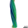
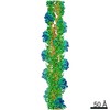
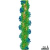
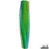
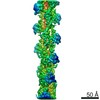
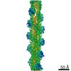
| Title | actin-like ParM protein bound to ADP | |||||||||
 Map data Map data | actin-like ParM protein bound to ADP | |||||||||
 Sample Sample |
| |||||||||
 Keywords Keywords |  bacterial cytoskeleton / plasmid segregation / actin-like protein bacterial cytoskeleton / plasmid segregation / actin-like protein | |||||||||
| Function / homology | Plasmid segregation protein ParM/StbA / : / Plasmid segregation protein ParM, N-terminal / Plasmid segregation protein ParM, C-terminal / ParM-like / plasmid partitioning / ATPase, nucleotide binding domain / identical protein binding / Plasmid segregation protein ParM Function and homology information Function and homology information | |||||||||
| Biological species |  Escherichia coli (E. coli) Escherichia coli (E. coli) | |||||||||
| Method | helical reconstruction / cryo EM / Resolution: 11.0 Å | |||||||||
 Authors Authors | Bharat TAM / Murshudov GN / Sachse C / Lowe J | |||||||||
 Citation Citation | Journal: Nature / Year: 2015 Title: Structures of actin-like ParM filaments show architecture of plasmid-segregating spindles. Authors: Tanmay A M Bharat / Garib N Murshudov / Carsten Sachse / Jan Löwe /   Abstract: Active segregation of Escherichia coli low-copy-number plasmid R1 involves formation of a bipolar spindle made of left-handed double-helical actin-like ParM filaments. ParR links the filaments with ...Active segregation of Escherichia coli low-copy-number plasmid R1 involves formation of a bipolar spindle made of left-handed double-helical actin-like ParM filaments. ParR links the filaments with centromeric parC plasmid DNA, while facilitating the addition of subunits to ParM filaments. Growing ParMRC spindles push sister plasmids to the cell poles. Here, using modern electron cryomicroscopy methods, we investigate the structures and arrangements of ParM filaments in vitro and in cells, revealing at near-atomic resolution how subunits and filaments come together to produce the simplest known mitotic machinery. To understand the mechanism of dynamic instability, we determine structures of ParM filaments in different nucleotide states. The structure of filaments bound to the ATP analogue AMPPNP is determined at 4.3 Å resolution and refined. The ParM filament structure shows strong longitudinal interfaces and weaker lateral interactions. Also using electron cryomicroscopy, we reconstruct ParM doublets forming antiparallel spindles. Finally, with whole-cell electron cryotomography, we show that doublets are abundant in bacterial cells containing low-copy-number plasmids with the ParMRC locus, leading to an asynchronous model of R1 plasmid segregation. | |||||||||
| History |
|
- Structure visualization
| Movie |
Movie viewer |
|---|---|
| Structure viewer | EM map: SurfViewMolmilJmol/JSmol |
| Supplemental images |

- Downloads & links
-EMDB archive
| Map data | emd_2848.map.gz | 492 KB | EMDB map data format | |
|---|---|---|---|---|
| Header (meta data) | emd-2848-v30.xmlemd-2848.xml | 8.7 KB 8.7 KB | Display Display | EMDB header |
| Images | 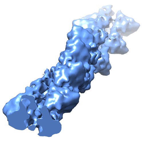 EMD-2848-parmadp.jpg EMD-2848-parmadp.jpg | 35.3 KB | ||
| Archive directory |  http://ftp.pdbj.org/pub/emdb/structures/EMD-2848ftp://ftp.pdbj.org/pub/emdb/structures/EMD-2848 http://ftp.pdbj.org/pub/emdb/structures/EMD-2848ftp://ftp.pdbj.org/pub/emdb/structures/EMD-2848 | HTTPS FTP |
-Related structure data
| Related structure data |  5ai7MUC 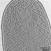 2849C  2850C  5aeyC M: atomic model generated by this map U: unfit; in different coordinate system*YM C: citing same article ( |
|---|---|
| Similar structure data |


-Links
| EMDB pages | EMDB (EBI/PDBe) / EMDataResource |
|---|
-Map
| File | Download / File: emd_2848.map.gz / Format: CCP4 / Size: 733.4 KB / Type: IMAGE STORED AS FLOATING POINT NUMBER (4 BYTES) | ||||||||||||||||||||||||||||||||||||||||||||||||||||||||||||
|---|---|---|---|---|---|---|---|---|---|---|---|---|---|---|---|---|---|---|---|---|---|---|---|---|---|---|---|---|---|---|---|---|---|---|---|---|---|---|---|---|---|---|---|---|---|---|---|---|---|---|---|---|---|---|---|---|---|---|---|---|---|
| Annotation | actin-like ParM protein bound to ADP | ||||||||||||||||||||||||||||||||||||||||||||||||||||||||||||
| Voxel size | X=Y=Z: 2.6 Å | ||||||||||||||||||||||||||||||||||||||||||||||||||||||||||||
| Density |
| ||||||||||||||||||||||||||||||||||||||||||||||||||||||||||||
| Symmetry | Space group: 1 | ||||||||||||||||||||||||||||||||||||||||||||||||||||||||||||
| Details | EMDB XML:
CCP4 map header:
| ||||||||||||||||||||||||||||||||||||||||||||||||||||||||||||
-Supplemental data
- Sample components
-Entire : ParM bound to ADP
| Entire | Name: ParM bound to ADP |
|---|---|
| Components |
|
-Supramolecule #1000: ParM bound to ADP
| Supramolecule | Name: ParM bound to ADP / type: sample / ID: 1000 / Details: ParM was incubated with ADP / Oligomeric state: filamentous / Number unique components: 1 |
|---|
-Macromolecule #1: ParM
| Macromolecule | Name: ParM / type: protein_or_peptide / ID: 1 / Details: ParM was incubated with ADP / Oligomeric state: Filamentous / Recombinant expression: Yes |
|---|---|
| Source (natural) | Organism: Escherichia coli (E. coli) |
| Molecular weight | Experimental: 36 KDa / Theoretical: 36 KDa |
| Recombinant expression | Organism: Escherichia coli BL21 (bacteria) / Recombinant cell: AI |
| Sequence | UniProtKB: Plasmid segregation protein ParM |
-Experimental details
-Structure determination
| Method | cryo EM |
|---|---|
 Processing Processing | helical reconstruction |
| Aggregation state | filament |
-Sample preparation
| Concentration | 14.4 mg/mL |
|---|---|
| Buffer | pH: 7 / Details: 50 mM Tris-HCl, 100 mM KCl, and 1 mM MgCl2 |
| Grid | Details: 200 mesh copper / rhodium grid with carbon support |
| Vitrification | Cryogen name: ETHANE / Chamber humidity: 100 % / Instrument: FEI VITROBOT MARK IV |
- Electron microscopy
| Microscope | FEI TITAN KRIOS |
|---|---|
| Electron beam | Acceleration voltage: 300 kV / Electron source: FIELD EMISSION GUN |
| Electron optics | Illumination mode: FLOOD BEAM / Imaging mode: BRIGHT FIELDBright-field microscopy / Cs: 2.7 mm / Nominal defocus max: -6.0 µm / Nominal defocus min: -2.0 µm |
| Sample stage | Specimen holder model: FEI TITAN KRIOS AUTOGRID HOLDER |
| Date | Jun 1, 2014 |
| Image recording | Category: CCD / Film or detector model: FEI FALCON II (4k x 4k) / Average electron dose: 25 e/Å2 |
| Experimental equipment |  Model: Titan Krios / Image courtesy: FEI Company |
-Image processing
| Final reconstruction | Applied symmetry - Helical parameters - Δz: 23.4 Å Applied symmetry - Helical parameters - Δ&Phi: 165.1 ° Applied symmetry - Helical parameters - Axial symmetry: C1 (asymmetric) Algorithm: OTHER / Resolution.type: BY AUTHOR / Resolution: 11.0 Å / Resolution method: OTHER / Software - Name: CTFFIND, EMAN2, SPRING |
|---|---|
| Details | CTF determination was conducted using CTFFIND. Data was extracted using EMAN2, and processed using SPRING. |
