 Movie
Movie Controller

[English] 日本語
 Yorodumi
- EMDB-26652: Prefusion-stabilized Nipah virus fusion protein complexed with Fab 4H3 -
+ Open data

- Basic information
| Entry | 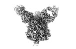 | |||||||||
|---|---|---|---|---|---|---|---|---|---|---|
| Title | Prefusion-stabilized Nipah virus fusion protein complexed with Fab 4H3 | |||||||||
 Map data Map data | Prefusion-stabilized Nipah virus fusion protein complexed with Fab 4H3 | |||||||||
 Sample Sample |
| |||||||||
 Keywords Keywords |  Henipavirus / Nipah virus / NiV / F / fusion / prefusion / preF / pre-F / neutralizing antibody / Fab / VIRAL PROTEIN / VIRAL PROTEIN-Immune System complex Henipavirus / Nipah virus / NiV / F / fusion / prefusion / preF / pre-F / neutralizing antibody / Fab / VIRAL PROTEIN / VIRAL PROTEIN-Immune System complex | |||||||||
| Function / homology |  Function and homology information Function and homology informationmembrane fusion involved in viral entry into host cell / symbiont entry into host cell / fusion of virus membrane with host plasma membrane / viral envelope / host cell plasma membrane / virion membrane / membraneSimilarity search - Function | |||||||||
| Biological species |  Nipah henipavirus / Nipah henipavirus /  Mus musculus (house mouse) Mus musculus (house mouse) | |||||||||
| Method | single particle reconstruction / cryo EM / Resolution: 2.8 Å | |||||||||
 Authors Authors | Byrne PO / McLellan JS | |||||||||
| Funding support |  United States, 1 items United States, 1 items
| |||||||||
 Citation Citation | Journal: Nat Commun / Year: 2023 Title: Structural basis for antibody recognition of vulnerable epitopes on Nipah virus F protein. Authors: Patrick O Byrne / Brian E Fisher / David R Ambrozak / Elizabeth G Blade / Yaroslav Tsybovsky / Barney S Graham / Jason S McLellan / Rebecca J Loomis /  Abstract: Nipah virus (NiV) is a pathogenic paramyxovirus that causes fatal encephalitis in humans. Two envelope glycoproteins, the attachment protein (G/RBP) and fusion protein (F), facilitate entry into host ...Nipah virus (NiV) is a pathogenic paramyxovirus that causes fatal encephalitis in humans. Two envelope glycoproteins, the attachment protein (G/RBP) and fusion protein (F), facilitate entry into host cells. Due to its vital role, NiV F presents an attractive target for developing vaccines and therapeutics. Several neutralization-sensitive epitopes on the NiV F apex have been described, however the antigenicity of most of the F protein's surface remains uncharacterized. Here, we immunize mice with prefusion-stabilized NiV F and isolate ten monoclonal antibodies that neutralize pseudotyped virus. Cryo-electron microscopy reveals eight neutralization-sensitive epitopes on NiV F, four of which have not previously been described. Novel sites span the lateral and basal faces of NiV F, expanding the known library of vulnerable epitopes. Seven of ten antibodies bind the Hendra virus (HeV) F protein. Multiple sequence alignment suggests that some of these newly identified neutralizing antibodies may also bind F proteins across the Henipavirus genus. This work identifies new epitopes as targets for therapeutics, provides a molecular basis for NiV neutralization, and lays a foundation for development of new cross-reactive antibodies targeting Henipavirus F proteins. | |||||||||
| History |
|
- Structure visualization
| Supplemental images |
|---|

- Downloads & links
-EMDB archive
| Map data | emd_26652.map.gz | 138.9 MB | EMDB map data format | |
|---|---|---|---|---|
| Header (meta data) | emd-26652-v30.xmlemd-26652.xml | 18.6 KB 18.6 KB | Display Display | EMDB header |
| FSC (resolution estimation) | emd_26652_fsc.xml | 12.1 KB | Display | FSC data file |
| Images |  emd_26652.png emd_26652.png | 53.4 KB | ||
| Masks | emd_26652_msk_1.map | 147.3 MB | Mask map | |
| Filedesc metadata | emd-26652.cif.gz | 5.9 KB | ||
| Others | emd_26652_additional_1.map.gzemd_26652_half_map_1.map.gzemd_26652_half_map_2.map.gz | 73.8 MB 136.9 MB 136.9 MB | ||
| Archive directory |  http://ftp.pdbj.org/pub/emdb/structures/EMD-26652ftp://ftp.pdbj.org/pub/emdb/structures/EMD-26652 http://ftp.pdbj.org/pub/emdb/structures/EMD-26652ftp://ftp.pdbj.org/pub/emdb/structures/EMD-26652 | HTTPS FTP |
-Related structure data
| Related structure data |  7uopMC  7up9C  7upaC 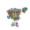 7upbC  7updC  7upkC M: atomic model generated by this map C: citing same article ( |
|---|---|
| Similar structure data |


-Links
| EMDB pages | EMDB (EBI/PDBe) / EMDataResource |
|---|---|
| Related items in Molecule of the Month |


-Map
| File | Download / File: emd_26652.map.gz / Format: CCP4 / Size: 147.3 MB / Type: IMAGE STORED AS FLOATING POINT NUMBER (4 BYTES) | ||||||||||||||||||||
|---|---|---|---|---|---|---|---|---|---|---|---|---|---|---|---|---|---|---|---|---|---|
| Annotation | Prefusion-stabilized Nipah virus fusion protein complexed with Fab 4H3 | ||||||||||||||||||||
| Voxel size | X=Y=Z: 1.1 Å | ||||||||||||||||||||
| Density |
| ||||||||||||||||||||
| Symmetry | Space group: 1 | ||||||||||||||||||||
| Details | EMDB XML:
|
-Supplemental data
-Mask #1
| File | emd_26652_msk_1.map | ||||||||||||
|---|---|---|---|---|---|---|---|---|---|---|---|---|---|
| Projections & Slices |
| ||||||||||||
| Density Histograms |
 Z
Z Y
Y X
X


-Additional map: Additional map 1
| File | emd_26652_additional_1.map | ||||||||||||
|---|---|---|---|---|---|---|---|---|---|---|---|---|---|
| Annotation | Additional map 1 | ||||||||||||
| Projections & Slices |
| ||||||||||||
| Density Histograms |


-Half map: Half Map 1
| File | emd_26652_half_map_1.map | ||||||||||||
|---|---|---|---|---|---|---|---|---|---|---|---|---|---|
| Annotation | Half Map 1 | ||||||||||||
| Projections & Slices |
| ||||||||||||
| Density Histograms |


-Half map: Half Map 2
| File | emd_26652_half_map_2.map | ||||||||||||
|---|---|---|---|---|---|---|---|---|---|---|---|---|---|
| Annotation | Half Map 2 | ||||||||||||
| Projections & Slices |
| ||||||||||||
| Density Histograms |


- Sample components
-Entire : Prefusion-stabilized Nipah virus fusion protein complexed with Fab 4H3
| Entire | Name: Prefusion-stabilized Nipah virus fusion protein complexed with Fab 4H3 |
|---|---|
| Components |
|
-Supramolecule #1: Prefusion-stabilized Nipah virus fusion protein complexed with Fab 4H3
| Supramolecule | Name: Prefusion-stabilized Nipah virus fusion protein complexed with Fab 4H3 type: complex / ID: 1 / Parent: 0 / Macromolecule list: #2-#3 |
|---|---|
| Source (natural) | Organism: Nipah henipavirus |
-Macromolecule #1: Fusion glycoprotein F0
| Macromolecule | Name: Fusion glycoprotein F0 / type: protein_or_peptide / ID: 1 / Number of copies: 3 / Enantiomer: LEVO |
|---|---|
| Source (natural) | Organism: Nipah henipavirus |
| Molecular weight | Theoretical: 52.295176 KDa |
| Recombinant expression | Organism:  Homo sapiens (human) Homo sapiens (human) |
| Sequence | String: MVVILDKRCY CNLLILILMI SECSVGILHY EKLSKIGLVK GVTRKYKIKS NPLTKDIVIK MIPNVSNMSQ CTGSVMENYK TRLNGILTP IKGALEIYKN NTHDCVGDVR LAGVCMAGVA IGIATAAQIT AGVALYEAMK NADNINKLKS SIESTNEAVV K LQETAEKT ...String: MVVILDKRCY CNLLILILMI SECSVGILHY EKLSKIGLVK GVTRKYKIKS NPLTKDIVIK MIPNVSNMSQ CTGSVMENYK TRLNGILTP IKGALEIYKN NTHDCVGDVR LAGVCMAGVA IGIATAAQIT AGVALYEAMK NADNINKLKS SIESTNEAVV K LQETAEKT VYVFTALQDY INTNLVPTID KIPCKQTELS LDLALSKYLS DLLFVFGPNL QDPVSNSMTI QAISQAFGGN YE TLLRTLG YATEDFDDLL ESDSITGQII YVDLSSYYII VRVYFPILTE IQQAYIQELL PVSFNNDNSE WISIVPNFIL VRN TLISNI EIGFCLITKR SVICNQDYAT PMTNNMRECL TGSTEKCPRE LVVSSHVPRF ALSNGVLFAN CISVTCQCQT TGRA ISQSG EQTLLMIDNT TCPTAVLGNV IISLGKYLGS VNYNSEGIAI GPPVFTDKVD ISSQISSMNQ SLQQSKDYIK UniProtKB: Fusion glycoprotein F0 |
-Macromolecule #2: Fab 4H3 heavy chain
| Macromolecule | Name: Fab 4H3 heavy chain / type: protein_or_peptide / ID: 2 / Number of copies: 3 / Enantiomer: LEVO |
|---|---|
| Source (natural) | Organism: Mus musculus (house mouse) |
| Molecular weight | Theoretical: 15.852915 KDa |
| Recombinant expression | Organism: Homo sapiens (human) |
| Sequence | String: MEFGLSWIFL AAILKGVQCQ IQLVQSGPEL KKPGETVKIS CKASGYTFRN YGVNWVKQGP GKDLKWMGWI NTLNGEPTYA DDFKRRFAF SLETSATTAF LQINNLKNED TATYFCARTF YDGYYYAMDY WGQGTSVTVS A |
-Macromolecule #3: Fab 4H3 light chain
| Macromolecule | Name: Fab 4H3 light chain / type: protein_or_peptide / ID: 3 / Number of copies: 3 / Enantiomer: LEVO |
|---|---|
| Source (natural) | Organism: Mus musculus (house mouse) |
| Molecular weight | Theoretical: 13.764521 KDa |
| Recombinant expression | Organism: Homo sapiens (human) |
| Sequence | String: MEFGLSWIFL AAILKGVQCE NVLTQSPTIM AASLGQKVTM TCSANSSVSS SYLHWYHQKS GASPKPLIHR TSNLASGVPA RFIGSGSGT SFSLTISSVE AEDDATYYCQ QWSGYPFITF GSGTKLEIK |
-Macromolecule #4: 2-acetamido-2-deoxy-beta-D-glucopyranose
| Macromolecule | Name: 2-acetamido-2-deoxy-beta-D-glucopyranose / type: ligand / ID: 4 / Number of copies: 12 / Formula: NAG |
|---|---|
| Molecular weight | Theoretical: 221.208 Da |
| Chemical component information |  ChemComp-NAG: |
-Experimental details
-Structure determination
| Method | cryo EM |
|---|---|
 Processing Processing | single particle reconstruction |
| Aggregation state | particle |
-Sample preparation
| Buffer | pH: 8 |
|---|---|
| Vitrification | Cryogen name: ETHANE |
- Electron microscopy
| Microscope | FEI TITAN KRIOS |
|---|---|
| Electron beam | Acceleration voltage: 300 kV / Electron source: FIELD EMISSION GUN |
| Electron optics | Illumination mode: FLOOD BEAM / Imaging mode: BRIGHT FIELDBright-field microscopy / Nominal defocus max: 2.5 µm / Nominal defocus min: 1.5 µm |
| Image recording | Film or detector model: GATAN K3 (6k x 4k) / Average electron dose: 80.0 e/Å2 |
| Experimental equipment |  Model: Titan Krios / Image courtesy: FEI Company |
-Image processing
| Startup model | Type of model: PDB ENTRY PDB model - PDB ID: |
|---|---|
| Initial angle assignment | Type: OTHER |
| Final angle assignment | Type: MAXIMUM LIKELIHOOD |
| Final reconstruction | Applied symmetry - Point group: C3 (3 fold cyclic) / Resolution.type: BY AUTHOR / Resolution: 2.8 Å / Resolution method: FSC 0.143 CUT-OFF / Number images used: 291096 |
| FSC plot (resolution estimation) |  |

