 Movie
Movie Controller


+ Open data

- Basic information
| Entry | Database: EMDB / ID: EMD-2610 | |||||||||
|---|---|---|---|---|---|---|---|---|---|---|
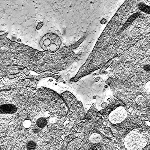
| Title | Electron tomography of zipping in Drosophyla melanogaster | |||||||||
 Map data Map data | Tomographic reconstruction of Drosophila zipping | |||||||||
 Sample Sample |
| |||||||||
 Keywords Keywords | epithelial tissue sealing / dorsal closure / serial tomography / high pressure freezing / freeze-substitution | |||||||||
| Biological species |  | |||||||||
| Method | electron tomography / cryo EM / negative staining | |||||||||
 Authors Authors | Eltsov M / Dube N / Yu Z / Haselmann-Weiss U / Brunner D / Frangakis A S | |||||||||
 Citation Citation | Journal: Nat Cell Biol / Year: 2015 Title: Quantitative analysis of cytoskeletal reorganization during epithelial tissue sealing by large-volume electron tomography. Authors: Mikhail Eltsov / Nadia Dubé / Zhou Yu / Laurynas Pasakarnis / Uta Haselmann-Weiss / Damian Brunner / Achilleas S Frangakis /    Abstract: The closure of epidermal openings is an essential biological process that causes major developmental problems such as spina bifida in humans if it goes awry. At present, the mechanism of closure ...The closure of epidermal openings is an essential biological process that causes major developmental problems such as spina bifida in humans if it goes awry. At present, the mechanism of closure remains elusive. Therefore, we reconstructed a model closure event, dorsal closure in fly embryos, by large-volume correlative electron tomography. We present a comprehensive, quantitative analysis of the cytoskeletal reorganization, enabling separated epidermal cells to seal the epithelium. After establishing contact through actin-driven exploratory filopodia, cells use a single lamella to generate 'roof tile'-like overlaps. These shorten to produce the force, 'zipping' the tissue closed. The shortening overlaps lack detectable actin filament ensembles but are crowded with microtubules. Cortical accumulation of shrinking microtubule ends suggests a force generation mechanism in which cortical motors pull on microtubule ends as for mitotic spindle positioning. In addition, microtubules orient filopodia and lamellae before zipping. Our 4D electron microscopy picture describes an entire developmental process and provides fundamental insight into epidermal closure. | |||||||||
| History |
|
- Structure visualization
| Movie |
 Movie viewer Movie viewer |
|---|---|
| Supplemental images |
- Downloads & links
-EMDB archive
| Map data | emd_2610.map.gz | 741.4 MB | EMDB map data format | |
|---|---|---|---|---|
| Header (meta data) | emd-2610-v30.xmlemd-2610.xml | 8.3 KB 8.3 KB | Display Display | EMDB header |
| Images | emd_2610.tif | 732.6 KB | ||
| Archive directory |  http://ftp.pdbj.org/pub/emdb/structures/EMD-2610ftp://ftp.pdbj.org/pub/emdb/structures/EMD-2610 http://ftp.pdbj.org/pub/emdb/structures/EMD-2610ftp://ftp.pdbj.org/pub/emdb/structures/EMD-2610 | HTTPS FTP |
-Validation report
| Summary document | emd_2610_validation.pdf.gz | 264.5 KB | Display | EMDB validaton report |
|---|---|---|---|---|
| Full document | emd_2610_full_validation.pdf.gz | 263.6 KB | Display | |
| Data in XML | emd_2610_validation.xml.gz | 4.9 KB | Display | |
| Arichive directory | https://ftp.pdbj.org/pub/emdb/validation_reports/EMD-2610ftp://ftp.pdbj.org/pub/emdb/validation_reports/EMD-2610 | HTTPS FTP |
-Links
| EMDB pages | EMDB (EBI/PDBe) / EMDataResource |
|---|
-Map
| File | Download / File: emd_2610.map.gz / Format: CCP4 / Size: 1 GB / Type: IMAGE STORED AS SIGNED BYTE | ||||||||||||||||||||||||||||||||||||||||||||||||||||||||||||
|---|---|---|---|---|---|---|---|---|---|---|---|---|---|---|---|---|---|---|---|---|---|---|---|---|---|---|---|---|---|---|---|---|---|---|---|---|---|---|---|---|---|---|---|---|---|---|---|---|---|---|---|---|---|---|---|---|---|---|---|---|---|
| Annotation | Tomographic reconstruction of Drosophila zipping | ||||||||||||||||||||||||||||||||||||||||||||||||||||||||||||
| Voxel size | X: 59 Å / Y: 59 Å / Z: 81 Å | ||||||||||||||||||||||||||||||||||||||||||||||||||||||||||||
| Density |
| ||||||||||||||||||||||||||||||||||||||||||||||||||||||||||||
| Symmetry | Space group: 1 | ||||||||||||||||||||||||||||||||||||||||||||||||||||||||||||
| Details | EMDB XML:
CCP4 map header:
| ||||||||||||||||||||||||||||||||||||||||||||||||||||||||||||
-Supplemental data
- Sample components
-Entire : Tomographic reconstruction of an entire zipping in Drosophila embryo
| Entire | Name: Tomographic reconstruction of an entire zipping in Drosophila embryo |
|---|---|
| Components |
|
-Supramolecule #1000: Tomographic reconstruction of an entire zipping in Drosophila embryo
| Supramolecule | Name: Tomographic reconstruction of an entire zipping in Drosophila embryo type: sample / ID: 1000 Details: Joined 34 serial tomograms were reconstructed from 2x2 montaged tilt-series. Tilt-series were recorded at x12000 corresponding to 0.9nm/pixel at the specimen level. The map was binned for ...Details: Joined 34 serial tomograms were reconstructed from 2x2 montaged tilt-series. Tilt-series were recorded at x12000 corresponding to 0.9nm/pixel at the specimen level. The map was binned for deposition to reduce the file size. The z-axis of the map corresponds to the anterior/posterior embryonic axis while the y-axis to the ventral/dorsal axis. The dorsal epidermal cells are located on the top. Number unique components: 1 |
|---|
-Supramolecule #1: zipping in Drosphila embryogenesis
| Supramolecule | Name: zipping in Drosphila embryogenesis / type: organelle_or_cellular_component / ID: 1 / Recombinant expression: No / Database: NCBI |
|---|---|
| Source (natural) | Organism: |
-Experimental details
-Structure determination
| Method | negative staining, cryo EM |
|---|---|
 Processing Processing | electron tomography |
| Aggregation state | tissue |
-Sample preparation
| Staining | Type: NEGATIVE Details: Sections were stained with 2% UA in 70% methanol and Reynolds lead citrate. |
|---|---|
| Grid | Details: slot grids with Formvar support |
| Vitrification | Cryogen name: NITROGEN / Instrument: OTHER |
- Electron microscopy
| Microscope | FEI TECNAI F30 |
|---|---|
| Date | May 10, 2010 |
| Image recording | Category: FILM / Film or detector model: FEI EAGLE (4k x 4k) / Digitization - Scanner: OTHER |
| Electron beam | Acceleration voltage: 300 kV / Electron source:  FIELD EMISSION GUN FIELD EMISSION GUN |
| Electron optics | Illumination mode: OTHER / Imaging mode: BRIGHT FIELD / Nominal magnification: 12000 |
| Sample stage | Specimen holder model: SIDE ENTRY, EUCENTRIC / Tilt series - Axis1 - Min angle: -60 ° / Tilt series - Axis1 - Max angle: 60 ° / Tilt series - Axis1 - Angle increment: 1.5 ° |
| Experimental equipment |  Model: Tecnai F30 / Image courtesy: FEI Company |
-Image processing
| Final reconstruction | Software - Name: ETOMO / Number images used: 120 |
|---|
