Movie
Movie Controller

[English] 日本語
 Yorodumi
- EMDB-2462: Structure and Host Adhesion Mechanism of Virulent Lactococcal Phage p2 -
+ Open data

- Basic information
| Entry | Database: EMDB / ID: EMD-2462 | |||||||||
|---|---|---|---|---|---|---|---|---|---|---|
| Title | Structure and Host Adhesion Mechanism of Virulent Lactococcal Phage p2 | |||||||||
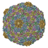
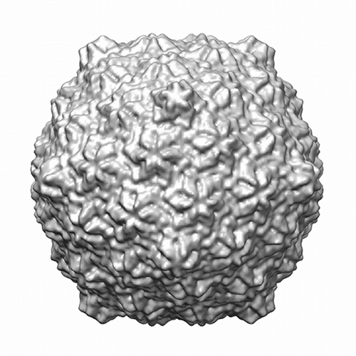
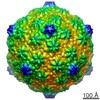
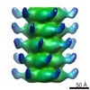
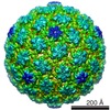
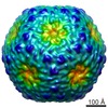
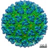
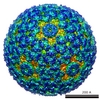
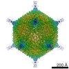
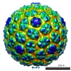
 Map data Map data | Icosahedral reconstruction of the capsid of lactococcal phage p2. | |||||||||
 Sample Sample |
| |||||||||
 Keywords Keywords |  Lactococcus lactis / Siphoviridae / 936 phages / Bacteriophage / electron microscopy / single-particle / p2 Lactococcus lactis / Siphoviridae / 936 phages / Bacteriophage / electron microscopy / single-particle / p2 | |||||||||
| Biological species |  Lactococcus lactis phage p2 (virus) Lactococcus lactis phage p2 (virus) | |||||||||
| Method | single particle reconstruction / cryo EM / Resolution: 13.0 Å | |||||||||
 Authors Authors | Bebeacua C / Tremblay D / Farenc C / Chapot MP / Sadovskaya I / van Heel M / Veesler D / Moineau S / Cambillau C | |||||||||
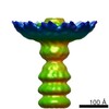
 Citation Citation | Journal: J Virol / Year: 2013 Title: Structure, adsorption to host, and infection mechanism of virulent lactococcal phage p2. Authors: Cecilia Bebeacua / Denise Tremblay / Carine Farenc / Marie-Pierre Chapot-Chartier / Irina Sadovskaya / Marin van Heel / David Veesler / Sylvain Moineau / Christian Cambillau /  Abstract: Lactococcal siphophages from the 936 and P335 groups infect the Gram-positive bacterium Lactococcus lactis using receptor binding proteins (RBPs) attached to their baseplate, a large multiprotein ...Lactococcal siphophages from the 936 and P335 groups infect the Gram-positive bacterium Lactococcus lactis using receptor binding proteins (RBPs) attached to their baseplate, a large multiprotein complex at the distal part of the tail. We have previously reported the crystal and electron microscopy (EM) structures of the baseplates of phages p2 (936 group) and TP901-1 (P335 group) as well as the full EM structure of the TP901-1 virion. Here, we report the complete EM structure of siphophage p2, including its capsid, connector complex, tail, and baseplate. Furthermore, we show that the p2 tail is characterized by the presence of protruding decorations, which are related to adhesins and are likely contributed by the major tail protein C-terminal domains. This feature is reminiscent of the tail of Escherichia coli phage λ and Bacillus subtilis phage SPP1 and might point to a common mechanism for establishing initial interactions with their bacterial hosts. Comparative analyses showed that the architecture of the phage p2 baseplate differs largely from that of lactococcal phage TP901-1. We quantified the interaction of its RBP with the saccharidic receptor and determined that specificity is due to lower k(off) values of the RBP/saccharidic dissociation. Taken together, these results suggest that the infection of L. lactis strains by phage p2 is a multistep process that involves reversible attachment, followed by baseplate activation, specific attachment of the RBPs to the saccharidic receptor, and DNA ejection. | |||||||||
| History |
|
- Structure visualization
| Movie |
 Movie viewer Movie viewer |
|---|---|
| Structure viewer | EM map: SurfViewMolmilJmol/JSmol |
| Supplemental images |

- Downloads & links
-EMDB archive
| Map data | emd_2462.map.gz | 5.6 MB | EMDB map data format | |
|---|---|---|---|---|
| Header (meta data) | emd-2462-v30.xmlemd-2462.xml | 10 KB 10 KB | Display Display | EMDB header |
| Images |  EMD-2462.png EMD-2462.png emd_2462.png emd_2462.png | 190.2 KB 190.2 KB | ||
| Archive directory |  http://ftp.pdbj.org/pub/emdb/structures/EMD-2462ftp://ftp.pdbj.org/pub/emdb/structures/EMD-2462 http://ftp.pdbj.org/pub/emdb/structures/EMD-2462ftp://ftp.pdbj.org/pub/emdb/structures/EMD-2462 | HTTPS FTP |
-Related structure data

-Links
| EMDB pages | EMDB (EBI/PDBe) / EMDataResource |
|---|
-Map
| File | Download / File: emd_2462.map.gz / Format: CCP4 / Size: 29.8 MB / Type: IMAGE STORED AS FLOATING POINT NUMBER (4 BYTES) | ||||||||||||||||||||||||||||||||||||||||||||||||||||||||||||||||||||
|---|---|---|---|---|---|---|---|---|---|---|---|---|---|---|---|---|---|---|---|---|---|---|---|---|---|---|---|---|---|---|---|---|---|---|---|---|---|---|---|---|---|---|---|---|---|---|---|---|---|---|---|---|---|---|---|---|---|---|---|---|---|---|---|---|---|---|---|---|---|
| Annotation | Icosahedral reconstruction of the capsid of lactococcal phage p2. | ||||||||||||||||||||||||||||||||||||||||||||||||||||||||||||||||||||
| Voxel size | X=Y=Z: 3.53 Å | ||||||||||||||||||||||||||||||||||||||||||||||||||||||||||||||||||||
| Density |
| ||||||||||||||||||||||||||||||||||||||||||||||||||||||||||||||||||||
| Symmetry | Space group: 1 | ||||||||||||||||||||||||||||||||||||||||||||||||||||||||||||||||||||
| Details | EMDB XML:
CCP4 map header:
| ||||||||||||||||||||||||||||||||||||||||||||||||||||||||||||||||||||
-Supplemental data
- Sample components
-Entire : Capsid of Lactococcal phage p2
| Entire | Name: Capsid of Lactococcal phage p2 |
|---|---|
| Components |
|
-Supramolecule #1000: Capsid of Lactococcal phage p2
| Supramolecule | Name: Capsid of Lactococcal phage p2 / type: sample / ID: 1000 / Oligomeric state: 60-mer / Number unique components: 1 |
|---|---|
| Molecular weight | Experimental: 3 MDa / Theoretical: 3 MDa |
-Supramolecule #1: Lactococcus lactis phage p2
| Supramolecule | Name: Lactococcus lactis phage p2 / type: virus / ID: 1 / Name.synonym: Lactococcal phage p2 Details: The capsid was selected from a sample containing entire Lactococcal phages p2. NCBI-ID: 100641 / Sci species name: Lactococcus lactis phage p2 / Virus type: OTHER / Virus isolate: SPECIES / Virus enveloped: No / Virus empty: No / Syn species name: Lactococcal phage p2 |
|---|---|
| Host (natural) | Organism: Lactococcal lactis / Strain: NZ9000 / synonym: BACTERIA(EUBACTERIA) |
| Virus shell | Shell ID: 1 / Name: T7 / Diameter: 660 Å / T number (triangulation number): 7 |
-Experimental details
-Structure determination
| Method | cryo EM |
|---|---|
 Processing Processing | single particle reconstruction |
| Aggregation state | particle |
-Sample preparation
| Buffer | pH: 7.5 / Details: 50 mM Tris-HCl, 100 mM NaCl, 8 mM MgSO4 |
|---|---|
| Grid | Details: Quantifoil grids |
| Vitrification | Cryogen name: ETHANE / Chamber humidity: 100 % / Instrument: FEI VITROBOT MARK I / Method: Blot for 2 seconds before plunging |
- Electron microscopy
| Microscope | FEI/PHILIPS CM200T |
|---|---|
| Electron beam | Acceleration voltage: 200 kV / Electron source: FIELD EMISSION GUN |
| Electron optics | Illumination mode: SPOT SCAN / Imaging mode: BRIGHT FIELDBright-field microscopy / Cs: 2.2 mm / Nominal defocus max: 0.002 µm / Nominal defocus min: 0.001 µm / Nominal magnification: 50000 |
| Sample stage | Specimen holder model: GATAN LIQUID NITROGEN |
| Alignment procedure | Legacy - Astigmatism: Objective lens astigmatism was corrected at 100,000 times magnification. |
| Date | Jul 1, 2009 |
| Image recording | Category: CCD / Film or detector model: GENERIC TVIPS (4k x 4k) / Digitization - Sampling interval: 3.53 µm / Number real images: 200 / Average electron dose: 10 e/Å2 |
-Image processing
| CTF correction | Details: Images |
|---|---|
| Final reconstruction | Applied symmetry - Point group: I (icosahedral) / Algorithm: OTHER / Resolution.type: BY AUTHOR / Resolution: 13.0 Å / Resolution method: OTHER / Software - Name: IMAGIC / Number images used: 3329 |
| Details | Reconstruction imposing Icosahedral reconstruction |
-Atomic model buiding 1
| Initial model | PDB ID: |
|---|---|
| Software | Name: Chimera |
| Details | 60 copies of the hexamer of HK97 MCP were manually fitted and automatically refined with Chimera |
| Refinement | Space: REAL / Protocol: RIGID BODY FIT |