Movie
Movie Controller

[English] 日本語
 Yorodumi
- EMDB-2441: Cryo-EM structure of activated and oligomeric restriction endonuc... -
+ Open data

- Basic information
| Entry | Database: EMDB / ID: EMD-2441 | |||||||||
|---|---|---|---|---|---|---|---|---|---|---|
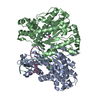
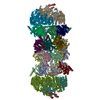
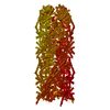
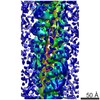
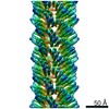
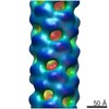
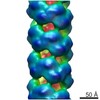
| Title | Cryo-EM structure of activated and oligomeric restriction endonuclease SgrAI | |||||||||
 Map data Map data | cryo-EM reconstruction of activated and oligomeric restriction endonuclease SgrAI | |||||||||
 Sample Sample |
| |||||||||
 Keywords Keywords |  restriction endonuclease / helix / oligomer / allostery / DNA cleavage / parasite-host interaction restriction endonuclease / helix / oligomer / allostery / DNA cleavage / parasite-host interaction | |||||||||
| Function / homology | Restriction endonuclease, type II, Cfr10I/Bse634I / Cfr10I/Bse634I restriction endonuclease / Restriction endonuclease type II-like / identical protein binding / metal ion binding / SgraIR restriction enzyme Function and homology information Function and homology information | |||||||||
| Biological species |  Streptomyces griseus (bacteria) Streptomyces griseus (bacteria) | |||||||||
| Method | single particle reconstruction / cryo EM / negative staining / Resolution: 8.6 Å | |||||||||
 Authors Authors | Lyumkis D / Talley H / Stewart A / Shah S / Park CK / Tama F / Potter CS / Carragher B / Horton NC | |||||||||
 Citation Citation | Journal: Structure / Year: 2013 Title: Allosteric regulation of DNA cleavage and sequence-specificity through run-on oligomerization. Authors: Dmitry Lyumkis / Heather Talley / Andrew Stewart / Santosh Shah / Chad K Park / Florence Tama / Clinton S Potter / Bridget Carragher / Nancy C Horton /  Abstract: SgrAI is a sequence specific DNA endonuclease that functions through an unusual enzymatic mechanism that is allosterically activated 200- to 500-fold by effector DNA, with a concomitant expansion of ...SgrAI is a sequence specific DNA endonuclease that functions through an unusual enzymatic mechanism that is allosterically activated 200- to 500-fold by effector DNA, with a concomitant expansion of its DNA sequence specificity. Using single-particle transmission electron microscopy to reconstruct distinct populations of SgrAI oligomers, we show that in the presence of allosteric, activating DNA, the enzyme forms regular, repeating helical structures characterized by the addition of DNA-binding dimeric SgrAI subunits in a run-on manner. We also present the structure of oligomeric SgrAI at 8.6 Å resolution, demonstrating the conformational state of SgrAI in its activated form. Activated and oligomeric SgrAI displays key protein-protein interactions near the helix axis between its N termini, as well as allosteric protein-DNA interactions that are required for enzymatic activation. The hybrid approach reveals an unusual mechanism of enzyme activation that explains SgrAI's oligomerization and allosteric behavior. | |||||||||
| History |
|
- Structure visualization
| Movie |
Movie viewer |
|---|---|
| Structure viewer | EM map: SurfViewMolmilJmol/JSmol |
| Supplemental images |

- Downloads & links
-EMDB archive
| Map data | emd_2441.map.gz | 22.4 MB | EMDB map data format | |
|---|---|---|---|---|
| Header (meta data) | emd-2441-v30.xmlemd-2441.xml | 13.9 KB 13.9 KB | Display Display | EMDB header |
| Images | 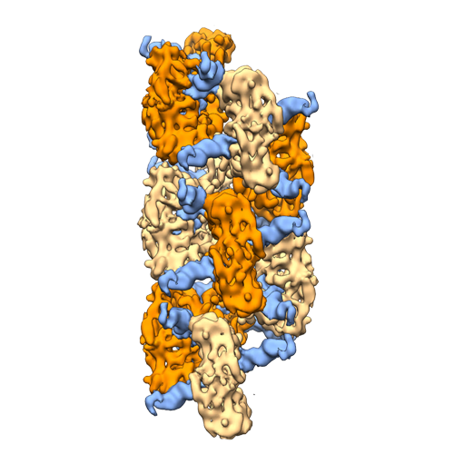 EMD-2441-EMDB_model_scale.png EMD-2441-EMDB_model_scale.png | 197.5 KB | ||
| Archive directory |  http://ftp.pdbj.org/pub/emdb/structures/EMD-2441ftp://ftp.pdbj.org/pub/emdb/structures/EMD-2441 http://ftp.pdbj.org/pub/emdb/structures/EMD-2441ftp://ftp.pdbj.org/pub/emdb/structures/EMD-2441 | HTTPS FTP |
-Related structure data
| Related structure data | 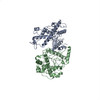 4c3gMC M: atomic model generated by this map C: citing same article ( |
|---|---|
| Similar structure data |


-Links
| EMDB pages | EMDB (EBI/PDBe) / EMDataResource |
|---|---|
| Related items in Molecule of the Month |


-Map
| File | Download / File: emd_2441.map.gz / Format: CCP4 / Size: 26.4 MB / Type: IMAGE STORED AS FLOATING POINT NUMBER (4 BYTES) | ||||||||||||||||||||||||||||||||||||||||||||||||||||||||||||||||||||
|---|---|---|---|---|---|---|---|---|---|---|---|---|---|---|---|---|---|---|---|---|---|---|---|---|---|---|---|---|---|---|---|---|---|---|---|---|---|---|---|---|---|---|---|---|---|---|---|---|---|---|---|---|---|---|---|---|---|---|---|---|---|---|---|---|---|---|---|---|---|
| Annotation | cryo-EM reconstruction of activated and oligomeric restriction endonuclease SgrAI | ||||||||||||||||||||||||||||||||||||||||||||||||||||||||||||||||||||
| Voxel size | X=Y=Z: 1.42 Å | ||||||||||||||||||||||||||||||||||||||||||||||||||||||||||||||||||||
| Density |
| ||||||||||||||||||||||||||||||||||||||||||||||||||||||||||||||||||||
| Symmetry | Space group: 1 | ||||||||||||||||||||||||||||||||||||||||||||||||||||||||||||||||||||
| Details | EMDB XML:
CCP4 map header:
| ||||||||||||||||||||||||||||||||||||||||||||||||||||||||||||||||||||
-Supplemental data
- Sample components
-Entire : cryo-EM reconstruction of activated and oligomeric restriction en...
| Entire | Name: cryo-EM reconstruction of activated and oligomeric restriction endonuclease SgrAI |
|---|---|
| Components |
|
-Supramolecule #1000: cryo-EM reconstruction of activated and oligomeric restriction en...
| Supramolecule | Name: cryo-EM reconstruction of activated and oligomeric restriction endonuclease SgrAI type: sample / ID: 1000 / Details: 1 megadalton (100 kDa) per DNA-bound dimer Oligomeric state: oligomeric SgrAI forms a helical array, whereby each asymmetric unit is composed of a DNA-bound SgrAI dimer Number unique components: 2 |
|---|---|
| Molecular weight | Theoretical: 1 MDa |
-Macromolecule #1: SgrAI
| Macromolecule | Name: SgrAI / type: protein_or_peptide / ID: 1 / Name.synonym: SgraIR restriction enzyme Details: 2 copies of pre-cleaved DNA containing sticky ends and the SgrAI recognition sequence is bound to an SgrAI dimer. The oligomeric helix is formed from multiple copies of such DNA-bound dimers Oligomeric state: DNA-bound dimer / Recombinant expression: Yes |
|---|---|
| Source (natural) | Organism: Streptomyces griseus (bacteria) |
| Molecular weight | Theoretical: 38 KDa |
| Recombinant expression | Organism: Escherichia coli (E. coli) / Recombinant strain: ER2566 / Recombinant plasmid: pET21a_SgrA1R |
| Sequence | UniProtKB: SgraIR restriction enzyme |
-Macromolecule #2: SgrAI recognition sequence
| Macromolecule | Name: SgrAI recognition sequence / type: dna / ID: 2 Details: 2 copies of pre-cleaved DNA containing sticky ends and the SgrAI recognition sequence is bound to an SgrAI dimer. The oligomeric helix is formed from multiple copies of such DNA-bound dimers Classification: DNA / Structure: DOUBLE HELIX / Synthetic?: No |
|---|---|
| Source (natural) | Organism: Streptomyces griseus (bacteria) |
| Sequence | String: GATGCGTGGG TCTTCACACC GGTGTGAAGA CCCACGCATC |
-Experimental details
-Structure determination
| Method | negative staining, cryo EM |
|---|---|
 Processing Processing | single particle reconstruction |
| Aggregation state | helical array |
-Sample preparation
| Concentration | 0.2 mg/mL |
|---|---|
| Buffer | pH: 8 Details: 10 mM Tris-HCl pH 8.0, 150 mM NaCl, 5 mM Mg(OAc)2, 0.5 mM DTT |
| Staining | Type: NEGATIVE Details: Specimens were prepared for cryo-EM by applying 3 microliters of sample in binding buffer to a holey carbon C-flat grid (CF-2/2-400) (Protochips, inc.) that had been plasma cleaned (Gatan, ...Details: Specimens were prepared for cryo-EM by applying 3 microliters of sample in binding buffer to a holey carbon C-flat grid (CF-2/2-400) (Protochips, inc.) that had been plasma cleaned (Gatan, Solarus) for 5 sec. The sample was allowed to adsorb to the grid for 30 sec., then plunge-frozen into liquid ethane using a manual cryo-plunger at 4 C. |
| Grid | Details: CF-2/2-400 |
| Vitrification | Cryogen name: ETHANE / Instrument: HOMEMADE PLUNGER Method: Specimens were prepared for cryo-EM by applying 3 microliters of sample in binding buffer to a holey carbon C-flat grid (CF-2/2-400 (Protochips, inc.) that had been plasma cleaned (Gatan, ...Method: Specimens were prepared for cryo-EM by applying 3 microliters of sample in binding buffer to a holey carbon C-flat grid (CF-2/2-400 (Protochips, inc.) that had been plasma cleaned (Gatan, Solarus) for 5 sec. The sample was allowed to adsorb to the grid for 30 sec., then plunge-frozen into liquid ethane using a manual cryo-plunger at 4 C. |
- Electron microscopy
| Microscope | FEI TECNAI F20 |
|---|---|
| Electron beam | Acceleration voltage: 200 kV / Electron source: FIELD EMISSION GUN |
| Electron optics | Calibrated magnification: 42134 / Illumination mode: FLOOD BEAM / Imaging mode: BRIGHT FIELDBright-field microscopy / Cs: 2 mm / Nominal defocus max: 3.0 µm / Nominal defocus min: 1.0 µm / Nominal magnification: 62000 |
| Sample stage | Specimen holder model: GATAN LIQUID NITROGEN |
| Alignment procedure | Legacy - Astigmatism: objective lens astigmatism was corrected at 120,000 times magnification |
| Date | Sep 19, 2012 |
| Image recording | Category: FILM / Film or detector model: DIRECT ELECTRON DE-12 (4k x 3k) / Digitization - Scanner: OTHER / Digitization - Sampling interval: 6.0 µm / Number real images: 655 / Average electron dose: 16 e/Å2 |
| Tilt angle min | 0 |
| Experimental equipment |  Model: Tecnai F20 / Image courtesy: FEI Company |
-Image processing
| CTF correction | Details: Frealign |
|---|---|
| Final reconstruction | Applied symmetry - Helical parameters - Δz: 21.6 Å Applied symmetry - Helical parameters - Δ&Phi: 86.2 ° Applied symmetry - Helical parameters - Axial symmetry: D1 (2x1 fold dihedral )Algorithm: OTHER / Resolution.type: BY AUTHOR / Resolution: 8.6 Å / Resolution method: FSC 0.143 CUT-OFF / Software - Name: Xmipp, and, Frealign / Number images used: 1918 |
| Details | The final reconstruction was obtained from 1,918 filament segments, averaged and inserted into the final reconstruction 8 times using Frealign |
-Atomic model buiding 1
| Initial model | PDB ID: Chain - #0 - Chain ID: A / Chain - #1 - Chain ID: B |
|---|---|
| Software | Name: Direx |
| Details | A model of SgrAI bound to PC DNA generated using the x-ray crystal structure of SgrAI bound to an 18 bp DNA containing a primary site (PDB ID: 3DVO) and additional modeled flanking DNA in B form was flexibly fitted using Direx, a geometry-based conformational sampling approach under low-resolution restraints. The refinement procedure was run for 500 steps followed by the minimization procedure of 300 steps |
| Refinement | Space: REAL / Protocol: FLEXIBLE FIT |
| Output model | PDB-4c3g: |