 Movie
Movie Controller

[English] 日本語
 Yorodumi
- EMDB-2410: Hsc70-induced Changes in Clathrin-Auxilin Cage Structure Suggest ... -
+ Open data

- Basic information
| Entry | Database: EMDB / ID: EMD-2410 | |||||||||
|---|---|---|---|---|---|---|---|---|---|---|
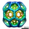
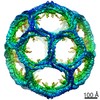
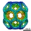
| Title | Hsc70-induced Changes in Clathrin-Auxilin Cage Structure Suggest a Role for Clathrin Light Chains in Cage Disassembly | |||||||||
 Map data Map data | Clathrin-auxilin-hsc70 complex | |||||||||
 Sample Sample |
| |||||||||
 Keywords Keywords |  Endocytosis / coated vesicles / clathrin / hsc70 / auxilin Endocytosis / coated vesicles / clathrin / hsc70 / auxilin | |||||||||
| Biological species |  Rattus (rat) / Bos taurus (cattle) Rattus (rat) / Bos taurus (cattle) | |||||||||
| Method | single particle reconstruction / cryo EM / Resolution: 34.0 Å | |||||||||
 Authors Authors | Young A / Stoilova-McPhie S / Rothnie A / Vallis Y / Harvey-Smith P / Ranson N / Kent H / Brodsky FM / Pearse BM / Roseman A / Smith CJ | |||||||||
 Citation Citation | Journal: Traffic / Year: 2013 Title: Hsc70-induced changes in clathrin-auxilin cage structure suggest a role for clathrin light chains in cage disassembly. Authors: Anna Young / Svetla Stoilova-McPhie / Alice Rothnie / Yvonne Vallis / Phillip Harvey-Smith / Neil Ranson / Helen Kent / Frances M Brodsky / Barbara M F Pearse / Alan Roseman / Corinne J Smith /  Abstract: The molecular chaperone, Hsc70, together with its co-factor, auxilin, facilitates the ATP-dependent removal of clathrin during clathrin-mediated endocytosis in cells. We have used cryo-electron ...The molecular chaperone, Hsc70, together with its co-factor, auxilin, facilitates the ATP-dependent removal of clathrin during clathrin-mediated endocytosis in cells. We have used cryo-electron microscopy to determine the 3D structure of a complex of clathrin, auxilin(401-910) and Hsc70 at pH 6 in the presence of ATP, frozen within 20 seconds of adding Hsc70 in order to visualize events that follow the binding of Hsc70 to clathrin and auxilin before clathrin disassembly. In this map, we observe density beneath the vertex of the cage that we attribute to bound Hsc70. This density emerges asymmetrically from the clathrin vertex, suggesting preferential binding by Hsc70 for one of the three possible sites at the vertex. Statistical comparison with a map of whole auxilin and clathrin previously published by us reveals the location of statistically significant differences which implicate involvement of clathrin light chains in structural rearrangements which occur after Hsc70 is recruited. Clathrin disassembly assays using light scattering suggest that loss of clathrin light chains reduces the efficiency with which auxilin facilitates this reaction. These data support a regulatory role for clathrin light chains in clathrin disassembly in addition to their established role in regulating clathrin assembly. | |||||||||
| History |
|
- Structure visualization
| Movie |
 Movie viewer Movie viewer |
|---|---|
| Structure viewer | EM map: SurfViewMolmilJmol/JSmol |
| Supplemental images |

- Downloads & links
-EMDB archive
| Map data | emd_2410.map.gz | 58.8 MB | EMDB map data format | |
|---|---|---|---|---|
| Header (meta data) | emd-2410-v30.xmlemd-2410.xml | 9 KB 9 KB | Display Display | EMDB header |
| Images | 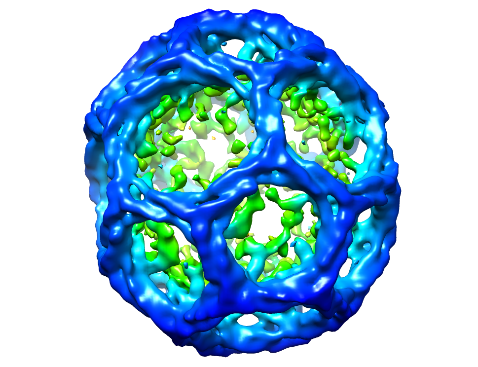 image.png image.png | 470.2 KB | ||
| Archive directory |  http://ftp.pdbj.org/pub/emdb/structures/EMD-2410ftp://ftp.pdbj.org/pub/emdb/structures/EMD-2410 http://ftp.pdbj.org/pub/emdb/structures/EMD-2410ftp://ftp.pdbj.org/pub/emdb/structures/EMD-2410 | HTTPS FTP |
-Related structure data

-Links
| EMDB pages | EMDB (EBI/PDBe) / EMDataResource |
|---|
-Map
| File | Download / File: emd_2410.map.gz / Format: CCP4 / Size: 62.5 MB / Type: IMAGE STORED AS FLOATING POINT NUMBER (4 BYTES) | ||||||||||||||||||||||||||||||||||||||||||||||||||||||||||||||||||||
|---|---|---|---|---|---|---|---|---|---|---|---|---|---|---|---|---|---|---|---|---|---|---|---|---|---|---|---|---|---|---|---|---|---|---|---|---|---|---|---|---|---|---|---|---|---|---|---|---|---|---|---|---|---|---|---|---|---|---|---|---|---|---|---|---|---|---|---|---|---|
| Annotation | Clathrin-auxilin-hsc70 complex | ||||||||||||||||||||||||||||||||||||||||||||||||||||||||||||||||||||
| Voxel size | X=Y=Z: 6.4 Å | ||||||||||||||||||||||||||||||||||||||||||||||||||||||||||||||||||||
| Density |
| ||||||||||||||||||||||||||||||||||||||||||||||||||||||||||||||||||||
| Symmetry | Space group: 1 | ||||||||||||||||||||||||||||||||||||||||||||||||||||||||||||||||||||
| Details | EMDB XML:
CCP4 map header:
| ||||||||||||||||||||||||||||||||||||||||||||||||||||||||||||||||||||
-Supplemental data
- Sample components
-Entire : Clathrin, auxilin (residues 401-910) and Hsc70
| Entire | Name: Clathrin, auxilin (residues 401-910) and Hsc70 |
|---|---|
| Components |
|
-Supramolecule #1000: Clathrin, auxilin (residues 401-910) and Hsc70
| Supramolecule | Name: Clathrin, auxilin (residues 401-910) and Hsc70 / type: sample / ID: 1000 / Oligomeric state: Not experimentally determined / Number unique components: 3 |
|---|
-Macromolecule #1: Hsc70
| Macromolecule | Name: Hsc70 / type: protein_or_peptide / ID: 1 / Recombinant expression: Yes |
|---|---|
| Source (natural) | Organism: Rattus (rat) / synonym: Rat |
-Macromolecule #2: auxilin401-910
| Macromolecule | Name: auxilin401-910 / type: protein_or_peptide / ID: 2 / Recombinant expression: Yes |
|---|---|
| Source (natural) | Organism: Bos taurus (cattle) / synonym: Cattle |
-Experimental details
-Structure determination
| Method | cryo EM |
|---|---|
 Processing Processing | single particle reconstruction |
| Aggregation state | particle |
-Sample preparation
| Buffer | pH: 6 Details: 20mM MES, 2mM magnesium acetate, 25mM KCl, 10mM (NH4)2SO4, 1.8mM Hepes, 18mM NaCl, 1mM DTT |
|---|---|
| Grid | Details: Quantifoil holey carbon grids, glow discharged in amylamine atmosphere |
| Vitrification | Cryogen name: ETHANE / Instrument: HOMEMADE PLUNGER Timed resolved state: Vitrified rapidly following addition of Hsc70-ATP |
- Electron microscopy
| Microscope | JEOL 2011 |
|---|---|
| Electron beam | Acceleration voltage: 200 kV / Electron source: LAB6 |
| Electron optics | Illumination mode: FLOOD BEAM / Imaging mode: BRIGHT FIELDBright-field microscopy / Nominal defocus max: 4.5 µm / Nominal defocus min: 1.0 µm / Nominal magnification: 40000 |
| Sample stage | Specimen holder model: GATAN LIQUID NITROGEN |
| Date | Jan 1, 2004 |
| Image recording | Category: FILM / Film or detector model: KODAK SO-163 FILM / Digitization - Scanner: NIKON COOLSCAN |
-Image processing
| CTF correction | Details: Whole micrographs |
|---|---|
| Final reconstruction | Applied symmetry - Point group: D6 (2x6 fold dihedral) / Algorithm: OTHER / Resolution.type: BY AUTHOR / Resolution: 34.0 Å / Resolution method: FSC 0.143 CUT-OFF / Software - Name: MRC, SPIDER, FREALIGN Details: The EM map has been sharpened to match a Fourier amplitude profile derived from a model of the hexagonal barrel (the biological unit) created from the crystal structure atomic model pdb ...Details: The EM map has been sharpened to match a Fourier amplitude profile derived from a model of the hexagonal barrel (the biological unit) created from the crystal structure atomic model pdb id1XI5. Fourier filtered to 30 Angstroms. Number images used: 1051 |
