 Movie
Movie Controller


+ Open data

- Basic information
| Entry | Database: EMDB / ID: EMD-2363 | |||||||||
|---|---|---|---|---|---|---|---|---|---|---|
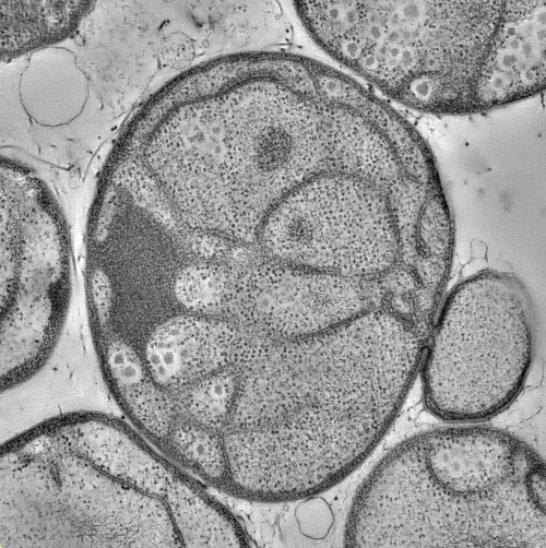
| Title | Electron tomogram through a Gemmata obscuriglobus cell | |||||||||
 Map data Map data | Tomographic reconstruction through a whole gemmata obscuriglobus cell. | |||||||||
 Sample Sample |
| |||||||||
 Keywords Keywords |  endomembrane / eukaryogenesis / electron tomography endomembrane / eukaryogenesis / electron tomography | |||||||||
| Biological species |  Gemmata obscuriglobus (bacteria) Gemmata obscuriglobus (bacteria) | |||||||||
| Method | electron tomography / cryo EM / negative staining | |||||||||
 Authors Authors | Santarella-Mellwig R / Pruggnaller S / Roos N / Mattaj IW / Devos DP | |||||||||
 Citation Citation | Journal: PLoS Biol / Year: 2013 Title: Three-dimensional reconstruction of bacteria with a complex endomembrane system. Authors: Rachel Santarella-Mellwig / Sabine Pruggnaller / Norbert Roos / Iain W Mattaj / Damien P Devos /  Abstract: The division of cellular space into functionally distinct membrane-defined compartments has been one of the major transitions in the history of life. Such compartmentalization has been claimed to ...The division of cellular space into functionally distinct membrane-defined compartments has been one of the major transitions in the history of life. Such compartmentalization has been claimed to occur in members of the Planctomycetes, Verrucomicrobiae, and Chlamydiae bacterial superphylum. Here we have investigated the three-dimensional organization of the complex endomembrane system in the planctomycete bacteria Gemmata obscuriglobus. We reveal that the G. obscuriglobus cells are neither compartmentalized nor nucleated as none of the spaces created by the membrane invaginations are closed; instead, they are all interconnected. Thus, the membrane organization of G. obscuriglobus, and most likely all PVC members, is not different from, but an extension of, the "classical" Gram-negative bacterial membrane system. Our results have implications for our definition and understanding of bacterial cell organization, the genesis of complex structure, and the origin of the eukaryotic endomembrane system. | |||||||||
| History |
|
- Structure visualization
| Movie |
 Movie viewer Movie viewer |
|---|---|
| Structure viewer | EM map: SurfViewMolmilJmol/JSmol |
| Supplemental images |
- Downloads & links
-EMDB archive
| Map data | emd_2363.map.gz | 2.6 GB | EMDB map data format | |
|---|---|---|---|---|
| Header (meta data) | emd-2363-v30.xmlemd-2363.xml | 6.9 KB 6.9 KB | Display Display | EMDB header |
| Images | EMD-2363-Gemmata_2363.tif | 764.5 KB | ||
| Others | g3d_model_11603.avi.gz | 35.3 MB | ||
| Archive directory |  http://ftp.pdbj.org/pub/emdb/structures/EMD-2363ftp://ftp.pdbj.org/pub/emdb/structures/EMD-2363 http://ftp.pdbj.org/pub/emdb/structures/EMD-2363ftp://ftp.pdbj.org/pub/emdb/structures/EMD-2363 | HTTPS FTP |
-Related structure data
-Links
| EMDB pages | EMDB (EBI/PDBe) / EMDataResource |
|---|
-Map
| File | Download / File: emd_2363.map.gz / Format: CCP4 / Size: 2.9 GB / Type: IMAGE STORED AS SIGNED BYTE | ||||||||||||||||||||||||||||||||||||||||||||||||||||||||||||||||||||
|---|---|---|---|---|---|---|---|---|---|---|---|---|---|---|---|---|---|---|---|---|---|---|---|---|---|---|---|---|---|---|---|---|---|---|---|---|---|---|---|---|---|---|---|---|---|---|---|---|---|---|---|---|---|---|---|---|---|---|---|---|---|---|---|---|---|---|---|---|---|
| Annotation | Tomographic reconstruction through a whole gemmata obscuriglobus cell. | ||||||||||||||||||||||||||||||||||||||||||||||||||||||||||||||||||||
| Voxel size | X=Y=Z: 15 Å | ||||||||||||||||||||||||||||||||||||||||||||||||||||||||||||||||||||
| Density |
| ||||||||||||||||||||||||||||||||||||||||||||||||||||||||||||||||||||
| Symmetry | Space group: 1 | ||||||||||||||||||||||||||||||||||||||||||||||||||||||||||||||||||||
| Details | EMDB XML:
CCP4 map header:
| ||||||||||||||||||||||||||||||||||||||||||||||||||||||||||||||||||||
-Supplemental data
-Others
- Sample components
-Entire : Gemmata obscuriglobus cell
| Entire | Name: Gemmata obscuriglobus cell |
|---|---|
| Components |
|
-Supramolecule #1000: Gemmata obscuriglobus cell
| Supramolecule | Name: Gemmata obscuriglobus cell / type: sample / ID: 1000 / Number unique components: 1 |
|---|
-Supramolecule #1: Gemmata obscuriglobus
| Supramolecule | Name: Gemmata obscuriglobus / type: organelle_or_cellular_component / ID: 1 / Recombinant expression: No / Database: NCBI |
|---|---|
| Source (natural) | Organism: Gemmata obscuriglobus (bacteria) / Strain: DSM-5831 |
-Experimental details
-Structure determination
| Method | negative staining, cryo EM |
|---|---|
 Processing Processing | electron tomography |
| Aggregation state | cell |
-Sample preparation
| Staining | Type: NEGATIVE Details: High pressure frozen/freeze substituted in 0.5% uranyl acetate |
|---|---|
| Vitrification | Cryogen name: NITROGEN / Instrument: OTHER Method: high pressure frozen with 100 micrometer deep carriers |
- Electron microscopy
| Microscope | FEI TECNAI F30 |
|---|---|
| Electron beam | Acceleration voltage: 300 kV / Electron source: FIELD EMISSION GUN |
| Electron optics | Illumination mode: FLOOD BEAM / Imaging mode: BRIGHT FIELDBright-field microscopy / Nominal magnification: 15500 |
| Sample stage | Specimen holder model: OTHER / Tilt series - Axis1 - Min angle: -60 ° / Tilt series - Axis1 - Max angle: 60 ° / Tilt series - Axis1 - Angle increment: 1 ° |
| Date | Jun 30, 2010 |
| Experimental equipment |  Model: Tecnai F30 / Image courtesy: FEI Company |
-Image processing
| Final reconstruction | Software - Name: IMOD / Number images used: 120 |
|---|
