 Movie
Movie Controller


+ Open data

- Basic information
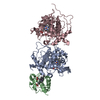
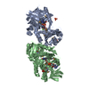
| Entry | Database: EMDB / ID: EMD-23482 | |||||||||
|---|---|---|---|---|---|---|---|---|---|---|
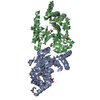
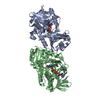
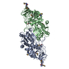
| Title | CryoEM structure of Escherichia coli PBP1b | |||||||||
 Map data Map data | LocScale sharpened final map. | |||||||||
 Sample Sample |
| |||||||||
 Keywords Keywords | Penicillin binding protein /  glycosyltransferase / transpeptidase / TRANSFERASE / HYDROLASE glycosyltransferase / transpeptidase / TRANSFERASE / HYDROLASE | |||||||||
| Function / homology |  Function and homology informationpeptidoglycan glycosyltransferase / positive regulation of bipolar cell growth / cell wall repair / peptidoglycan glycosyltransferase activity / serine-type D-Ala-D-Ala carboxypeptidase / serine-type D-Ala-D-Ala carboxypeptidase activity / penicillin binding / peptidoglycan biosynthetic process / peptidoglycan-based cell wall / regulation of cell shape ...peptidoglycan glycosyltransferase / positive regulation of bipolar cell growth / cell wall repair / peptidoglycan glycosyltransferase activity / serine-type D-Ala-D-Ala carboxypeptidase / serine-type D-Ala-D-Ala carboxypeptidase activity / penicillin binding / peptidoglycan biosynthetic process / peptidoglycan-based cell wall / regulation of cell shape / outer membrane-bounded periplasmic space / response to antibiotic / proteolysis / membrane / plasma membrane Function and homology informationpeptidoglycan glycosyltransferase / positive regulation of bipolar cell growth / cell wall repair / peptidoglycan glycosyltransferase activity / serine-type D-Ala-D-Ala carboxypeptidase / serine-type D-Ala-D-Ala carboxypeptidase activity / penicillin binding / peptidoglycan biosynthetic process / peptidoglycan-based cell wall / regulation of cell shape ...peptidoglycan glycosyltransferase / positive regulation of bipolar cell growth / cell wall repair / peptidoglycan glycosyltransferase activity / serine-type D-Ala-D-Ala carboxypeptidase / serine-type D-Ala-D-Ala carboxypeptidase activity / penicillin binding / peptidoglycan biosynthetic process / peptidoglycan-based cell wall / regulation of cell shape / outer membrane-bounded periplasmic space / response to antibiotic / proteolysis / membrane / plasma membraneSimilarity search - Function | |||||||||
| Biological species |  Escherichia coli (E. coli) / Escherichia coli (strain K12) (bacteria) Escherichia coli (E. coli) / Escherichia coli (strain K12) (bacteria) | |||||||||
| Method | single particle reconstruction / cryo EM / Resolution: 3.28 Å | |||||||||
 Authors Authors | Caveney NA / Workman SD | |||||||||
| Funding support |  Canada, 1 items Canada, 1 items
| |||||||||
 Citation Citation | Journal: Nat Commun / Year: 2021 Title: CryoEM structure of the antibacterial target PBP1b at 3.3 Å resolution. Authors: Nathanael A Caveney / Sean D Workman / Rui Yan / Claire E Atkinson / Zhiheng Yu / Natalie C J Strynadka /  Abstract: The pathway for the biosynthesis of the bacterial cell wall is one of the most prolific antibiotic targets, exemplified by the widespread use of β-lactam antibiotics. Despite this, our structural ...The pathway for the biosynthesis of the bacterial cell wall is one of the most prolific antibiotic targets, exemplified by the widespread use of β-lactam antibiotics. Despite this, our structural understanding of class A penicillin binding proteins, which perform the last two steps in this pathway, is incomplete due to the inherent difficulty in their crystallization and the complexity of their substrates. Here, we determine the near atomic resolution structure of the 83 kDa class A PBP from Escherichia coli, PBP1b, using cryogenic electron microscopy and a styrene maleic acid anhydride membrane mimetic. PBP1b, in its apo form, is seen to exhibit a distinct conformation in comparison to Moenomycin-bound crystal structures. The work herein paves the way for the use of cryoEM in structure-guided antibiotic development for this notoriously difficult to crystalize class of proteins and their complex substrates. | |||||||||
| History |
|
- Structure visualization
| Movie |
Movie viewer |
|---|---|
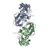
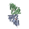
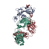
| Structure viewer | EM map: SurfViewMolmilJmol/JSmol |
| Supplemental images |

- Downloads & links
-EMDB archive
| Map data | emd_23482.map.gz | 3.2 MB | EMDB map data format | |
|---|---|---|---|---|
| Header (meta data) | emd-23482-v30.xmlemd-23482.xml | 15.1 KB 15.1 KB | Display Display | EMDB header |
| FSC (resolution estimation) | emd_23482_fsc.xml | 13.7 KB | Display | FSC data file |
| Images | 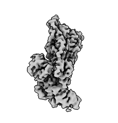 emd_23482.png emd_23482.png | 35.7 KB | ||
| Filedesc metadata | emd-23482.cif.gz | 5.6 KB | ||
| Others | emd_23482_half_map_1.map.gzemd_23482_half_map_2.map.gz | 169.8 MB 169.7 MB | ||
| Archive directory |  http://ftp.pdbj.org/pub/emdb/structures/EMD-23482ftp://ftp.pdbj.org/pub/emdb/structures/EMD-23482 http://ftp.pdbj.org/pub/emdb/structures/EMD-23482ftp://ftp.pdbj.org/pub/emdb/structures/EMD-23482 | HTTPS FTP |
-Related structure data
| Related structure data |  7lq6MC M: atomic model generated by this map C: citing same article ( |
|---|---|
| Similar structure data |

-Links
| EMDB pages | EMDB (EBI/PDBe) / EMDataResource |
|---|---|
| Related items in Molecule of the Month |

-Map
| File | Download / File: emd_23482.map.gz / Format: CCP4 / Size: 216 MB / Type: IMAGE STORED AS FLOATING POINT NUMBER (4 BYTES) | ||||||||||||||||||||||||||||||||||||||||||||||||||||||||||||||||||||
|---|---|---|---|---|---|---|---|---|---|---|---|---|---|---|---|---|---|---|---|---|---|---|---|---|---|---|---|---|---|---|---|---|---|---|---|---|---|---|---|---|---|---|---|---|---|---|---|---|---|---|---|---|---|---|---|---|---|---|---|---|---|---|---|---|---|---|---|---|---|
| Annotation | LocScale sharpened final map. | ||||||||||||||||||||||||||||||||||||||||||||||||||||||||||||||||||||
| Voxel size | X=Y=Z: 0.844 Å | ||||||||||||||||||||||||||||||||||||||||||||||||||||||||||||||||||||
| Density |
| ||||||||||||||||||||||||||||||||||||||||||||||||||||||||||||||||||||
| Symmetry | Space group: 1 | ||||||||||||||||||||||||||||||||||||||||||||||||||||||||||||||||||||
| Details | EMDB XML:
CCP4 map header:
| ||||||||||||||||||||||||||||||||||||||||||||||||||||||||||||||||||||
-Supplemental data
-Half map: Half-map 2.
| File | emd_23482_half_map_1.map | ||||||||||||
|---|---|---|---|---|---|---|---|---|---|---|---|---|---|
| Annotation | Half-map 2. | ||||||||||||
| Projections & Slices |
| ||||||||||||
| Density Histograms |
 Z
Z Y
Y X
X


-Half map: Half-map 1.
| File | emd_23482_half_map_2.map | ||||||||||||
|---|---|---|---|---|---|---|---|---|---|---|---|---|---|
| Annotation | Half-map 1. | ||||||||||||
| Projections & Slices |
| ||||||||||||
| Density Histograms |


- Sample components
-Entire : Escherichia coli PBP1b in SMA
| Entire | Name: Escherichia coli PBP1b in SMA |
|---|---|
| Components |
|
-Supramolecule #1: Escherichia coli PBP1b in SMA
| Supramolecule | Name: Escherichia coli PBP1b in SMA / type: complex / ID: 1 / Parent: 0 / Macromolecule list: all |
|---|---|
| Source (natural) | Organism: Escherichia coli (E. coli) |
| Molecular weight | Theoretical: 83 KDa |
-Macromolecule #1: Penicillin-binding protein 1B
| Macromolecule | Name: Penicillin-binding protein 1B / type: protein_or_peptide / ID: 1 / Number of copies: 1 / Enantiomer: LEVO / EC number: peptidoglycan glycosyltransferase |
|---|---|
| Source (natural) | Organism: Escherichia coli (strain K12) (bacteria) / Strain: K12 |
| Molecular weight | Theoretical: 83.280352 KDa |
| Recombinant expression | Organism: Escherichia coli (E. coli) |
| Sequence | String: KPRGKRGWLW LLLKLAIVFA VLIAIYGVYL DQKIRSRIDG KVWQLPAAVY GRMVNLEPDM TISKNEMVKL LEATQYRQVS KMTRPGEFT VQANSIEMIR RPFDFPDSKE GQVRARLTFD GDHLATIVNM ENNRQFGFFR LDPRLITMIS SPNGEQRLFV P RSGFPDLL ...String: KPRGKRGWLW LLLKLAIVFA VLIAIYGVYL DQKIRSRIDG KVWQLPAAVY GRMVNLEPDM TISKNEMVKL LEATQYRQVS KMTRPGEFT VQANSIEMIR RPFDFPDSKE GQVRARLTFD GDHLATIVNM ENNRQFGFFR LDPRLITMIS SPNGEQRLFV P RSGFPDLL VDTLLATEDR HFYEHDGISL YSIGRAVLAN LTAGRTVQGA STLTQQLVKN LFLSSERSYW RKANEAYMAL IM DARYSKD RILELYMNEV YLGQSGDNEI RGFPLASLYY FGRPVEELSL DQQALLVGMV KGASIYNPWR NPKLALERRN LVL RLLQQQ QIIDQELYDM LSARPLGVQP RGGVISPQPA FMQLVRQELQ AKLGDKVKDL SGVKIFTTFD SVAQDAAEKA AVEG IPALK KQRKLSDLET AIVVVDRFSG EVRAMVGGSE PQFAGYNRAM QARRSIGSLA KPATYLTALS QPKIYRLNTW IADAP IALR QPNGQVWSPQ NDDRRYSESG RVMLVDALTR SMNVPTVNLG MALGLPAVTE TWIKLGVPKD QLHPVPAMLL GALNLT PIE VAQAFQTIAS GGNRAPLSAL RSVIAEDGKV LYQSFPQAER AVPAQAAYLT LWTMQQVVQR GTGRQLGAKY PNLHLAG KT GTTNNNVDTW FAGIDGSTVT ITWVGRDNNQ PTKLYGASGA MSIYQRYLAN QTPTPLNLVP PEDIADMGVD YDGNFVCS G GMRILPVWTS DPQSLCQQSE MQQQPS UniProtKB: Penicillin-binding protein 1B |
-Experimental details
-Structure determination
| Method | cryo EM |
|---|---|
 Processing Processing | single particle reconstruction |
| Aggregation state | particle |
-Sample preparation
| Concentration | 0.125 mg/mL |
|---|---|
| Buffer | pH: 8 |
| Grid | Model: Quantifoil R1.2/1.3 / Material: COPPER / Mesh: 300 / Pretreatment - Type: GLOW DISCHARGE |
| Vitrification | Cryogen name: ETHANE / Chamber humidity: 100 % / Chamber temperature: 277.15 K / Instrument: FEI VITROBOT MARK IV / Details: 3 blot force, 3 second blot. |
- Electron microscopy
| Microscope | TFS KRIOS |
|---|---|
| Electron beam | Acceleration voltage: 300 kV / Electron source: FIELD EMISSION GUN |
| Electron optics | Calibrated magnification: 105000 / Illumination mode: OTHER / Imaging mode: OTHER / Cs: 2.7 mm |
| Sample stage | Specimen holder model: FEI TITAN KRIOS AUTOGRID HOLDER / Cooling holder cryogen: NITROGEN |
| Image recording | Film or detector model: GATAN K3 (6k x 4k) / Average electron dose: 60.0 e/Å2 |
| Experimental equipment |  Model: Titan Krios / Image courtesy: FEI Company |
-Image processing
| Startup model | Type of model: INSILICO MODEL |
|---|---|
| Initial angle assignment | Type: OTHER |
| Final angle assignment | Type: OTHER |
| Final reconstruction | Resolution.type: BY AUTHOR / Resolution: 3.28 Å / Resolution method: FSC 0.143 CUT-OFF / Software: (Name: RELION, cryoSPARC) / Number images used: 462997 |
| FSC plot (resolution estimation) |  |
