 Movie
Movie Controller


+ Open data

- Basic information
| Entry | Database: EMDB / ID: EMD-2328 | |||||||||
|---|---|---|---|---|---|---|---|---|---|---|
| Title | Structure of the bacterial V-ATPase from Thermus thermophilius | |||||||||
 Map data Map data | Reconstruction of the bacterial V-ATPase | |||||||||
 Sample Sample |
| |||||||||
 Keywords Keywords | bacterial V-ATPase / rotary ATPase | |||||||||
| Biological species |    Thermus thermophilus (bacteria) Thermus thermophilus (bacteria) | |||||||||
| Method | electron crystallography / negative staining / Resolution: 18.0 Å | |||||||||
 Authors Authors | Tani K / Arthur CP / Tamakoshi M / Yokoyama K / Mitsuoka K / Fujiyoshi Y / Gerle C | |||||||||
 Citation Citation | Journal: Microscopy (Oxf) / Year: 2013 Title: Visualization of two distinct states of disassembly in the bacterial V-ATPase from Thermus thermophilus. Authors: Kazutoshi Tani / Christopher P Arthur / Masatada Tamakoshi / Ken Yokoyama / Kaoru Mitsuoka / Yoshinori Fujiyoshi / Christoph Gerle /  Abstract: V-ATPases are multisubunit, membrane-bound, energy-converting, cellular machines whose assembly and disassembly is innately connected to their activity in vivo. In vitro V-ATPases show a propensity ...V-ATPases are multisubunit, membrane-bound, energy-converting, cellular machines whose assembly and disassembly is innately connected to their activity in vivo. In vitro V-ATPases show a propensity for disassembly that greatly complicates their functional, and, in particular, structural characterization. Direct structural evidence for early stages of their disassembly has not been reported yet. We analyzed the structure of the V-ATPase from Thermus thermophilus in a single negatively stained two-dimensional (2-D) crystal both by electron tomography and by electron crystallography. Our analysis demonstrated that for 2-D crystals of fragile macromolecular complexes, which are too heterogenous or too few for the merging of image data from many crystals, single-crystal 3-D reconstructions by electron tomography and electron crystallography are expedient tools of analysis. The asymmetric unit in the 2-D crystal lattice contains two different V-ATPase complexes that appear to be in an early stage of disassembly and with either one or both peripheral stalks not being visualized, suggesting the involvement of the peripheral stalks in early stages of disassembly. | |||||||||
| History |
|
- Structure visualization
| Movie |
 Movie viewer Movie viewer |
|---|---|
| Structure viewer | EM map: SurfViewMolmilJmol/JSmol |
| Supplemental images |


- Downloads & links
-EMDB archive
| Map data | emd_2328.map.gz | 424.9 KB | EMDB map data format | |
|---|---|---|---|---|
| Header (meta data) | emd-2328-v30.xmlemd-2328.xml | 9.5 KB 9.5 KB | Display Display | EMDB header |
| Images | 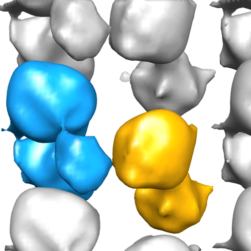 2328_emd-2328-VA_emdep.jpg 2328_emd-2328-VA_emdep.jpg emd-2328-VA_emdep.jpg emd-2328-VA_emdep.jpg | 132.5 KB 132.5 KB | ||
| Archive directory |  http://ftp.pdbj.org/pub/emdb/structures/EMD-2328ftp://ftp.pdbj.org/pub/emdb/structures/EMD-2328 http://ftp.pdbj.org/pub/emdb/structures/EMD-2328ftp://ftp.pdbj.org/pub/emdb/structures/EMD-2328 | HTTPS FTP |
-Links
| EMDB pages | EMDB (EBI/PDBe) / EMDataResource |
|---|
-Map
| File | Download / File: emd_2328.map.gz / Format: CCP4 / Size: 476.6 KB / Type: IMAGE STORED AS FLOATING POINT NUMBER (4 BYTES) | ||||||||||||||||||||||||||||||||||||||||||||||||||||||||||||||||||||
|---|---|---|---|---|---|---|---|---|---|---|---|---|---|---|---|---|---|---|---|---|---|---|---|---|---|---|---|---|---|---|---|---|---|---|---|---|---|---|---|---|---|---|---|---|---|---|---|---|---|---|---|---|---|---|---|---|---|---|---|---|---|---|---|---|---|---|---|---|---|
| Annotation | Reconstruction of the bacterial V-ATPase | ||||||||||||||||||||||||||||||||||||||||||||||||||||||||||||||||||||
| Voxel size | X: 4.296 Å / Y: 4.4 Å / Z: 4.166 Å | ||||||||||||||||||||||||||||||||||||||||||||||||||||||||||||||||||||
| Density |
| ||||||||||||||||||||||||||||||||||||||||||||||||||||||||||||||||||||
| Symmetry | Space group: 1 | ||||||||||||||||||||||||||||||||||||||||||||||||||||||||||||||||||||
| Details | EMDB XML:
CCP4 map header:
| ||||||||||||||||||||||||||||||||||||||||||||||||||||||||||||||||||||
-Supplemental data
- Sample components
-Entire : Bacterial V-type ATPase
| Entire | Name: Bacterial V-type ATPase |
|---|---|
| Components |
|
-Supramolecule #1000: Bacterial V-type ATPase
| Supramolecule | Name: Bacterial V-type ATPase / type: sample / ID: 1000 / Number unique components: 1 |
|---|
-Macromolecule #1: V-ATPase
| Macromolecule | Name: V-ATPase / type: protein_or_peptide / ID: 1 / Number of copies: 2 / Recombinant expression: No / Database: NCBI |
|---|---|
| Source (natural) | Organism: Thermus thermophilus (bacteria) / Location in cell: Plasma membrane |
-Experimental details
-Structure determination
| Method | negative staining |
|---|---|
 Processing Processing | electron crystallography |
| Aggregation state | 2D array |
-Sample preparation
| Concentration | 1 mg/mL |
|---|---|
| Staining | Type: NEGATIVE Details: A 2.5 ul sample was transferred to a glow-discharged carbon coated copper grid. After 1 min, the drop was blotted with a slice of filter paper, and then the grid was washed with 2.5 ul water ...Details: A 2.5 ul sample was transferred to a glow-discharged carbon coated copper grid. After 1 min, the drop was blotted with a slice of filter paper, and then the grid was washed with 2.5 ul water to avoid the precipitation caused by mixing phosphate buffer with uranyl acetate. Subsequently, the sample was stained with 2.5 ul of 2% uranyl acetate and air-dried. |
| Grid | Details: a glow-discharged carbon coated copper grid |
| Vitrification | Cryogen name: NONE / Instrument: OTHER |
| Details | Crystals grown by dialysis |
| Crystal formation | Details: Crystals grown by dialysis |
- Electron microscopy
| Microscope | FEI TECNAI F30 |
|---|---|
| Electron beam | Acceleration voltage: 300 kV / Electron source: FIELD EMISSION GUN |
| Electron optics | Illumination mode: FLOOD BEAM / Imaging mode: BRIGHT FIELDBright-field microscopy / Nominal defocus max: 2.27 µm / Nominal defocus min: 1.4 µm / Nominal magnification: 40000 |
| Sample stage | Specimen holder model: SIDE ENTRY, EUCENTRIC / Tilt angle min: -55 / Tilt angle max: 55 / Tilt series - Axis1 - Min angle: -55 ° / Tilt series - Axis1 - Max angle: 55 ° |
| Date | Nov 28, 2007 |
| Image recording | Category: CCD / Film or detector model: GENERIC GATAN / Digitization - Sampling interval: 15 µm / Number real images: 212 Details: Every image was recorded by 2Kx2K CCD camera (Gatan) Bits/pixel: 16 |
| Experimental equipment |  Model: Tecnai F30 / Image courtesy: FEI Company |
-Image processing
| Crystal parameters | Unit cell - A: 232 Å / Unit cell - B: 132 Å / Unit cell - C: 300 Å / Unit cell - γ: 90.0 ° / Unit cell - α: 90.0 ° / Unit cell - β: 90.0 ° / Plane group: P 1 |
|---|---|
| Final reconstruction | Resolution.type: BY AUTHOR / Resolution: 18.0 Å / Resolution method: OTHER / Software - Name: MRC |
| Details | Images were processed using MRC package |
