- EMDB-2321: The bacterial DnaC helicase loader is a DnaB ring breaker -
+
Open data
ID or keywords:
Loading...
-
Basic information
Entry
Database: EMDB / ID: EMD-2321
Title
The bacterial DnaC helicase loader is a DnaB ring breaker
Map data
Negative staining reconstruction of E. coli DnaB/DnaC complex
Sample
Sample: E. coli DnaB helicase bound to the DnaC loading factor
Protein or peptide: DnaB replicative helicase
Protein or peptide: DnaC helicase loading factor
Keywords
Bacterial DNA replication / DNA replication initiation / helicase / helicase loader / DnaB / DnaC / electron microscopy / SAXS / clamp loader / structure
Function / homology
Function and homology information
DnaB-DnaC complex / DnaB-DnaC-Rep-PriC complex / DnaB-DnaG complex / DnaB-DnaC-DnaT-PriA-PriC complex / DNA helicase complex / DnaB-DnaC-DnaT-PriA-PriB complex / primosome complex / replisome / DNA replication, synthesis of primer / DNA strand elongation involved in DNA replication ...DnaB-DnaC complex / DnaB-DnaC-Rep-PriC complex / DnaB-DnaG complex / DnaB-DnaC-DnaT-PriA-PriC complex / DNA helicase complex / DnaB-DnaC-DnaT-PriA-PriB complex / primosome complex / replisome / DNA replication, synthesis of primer / DNA strand elongation involved in DNA replication / DNA duplex unwinding / response to ionizing radiation / replication fork processing / DNA unwinding involved in DNA replication / DNA replication initiation / DNA helicase activity / helicase activity / DNA helicase / DNA replication / ATP hydrolysis activity / DNA binding / ATP binding / identical protein binding / cytosol Similarity search - Function
DNA replication protein DnaC/insertion sequence putative ATP-binding protein / IstB-like ATP-binding protein / IstB-like ATP binding protein / DNA helicase, DnaB type / DNA helicase, DnaB type / DNA helicase, DnaB-like, N-terminal / DnaB-like helicase N terminal domain / DNA helicase, DnaB-like, N-terminal domain superfamily / DNA helicase DnaB, N-terminal/DNA primase DnaG, C-terminal / DnaB-like helicase C terminal domain ...DNA replication protein DnaC/insertion sequence putative ATP-binding protein / IstB-like ATP-binding protein / IstB-like ATP binding protein / DNA helicase, DnaB type / DNA helicase, DnaB type / DNA helicase, DnaB-like, N-terminal / DnaB-like helicase N terminal domain / DNA helicase, DnaB-like, N-terminal domain superfamily / DNA helicase DnaB, N-terminal/DNA primase DnaG, C-terminal / DnaB-like helicase C terminal domain / DNA helicase, DnaB-like, C-terminal / Superfamily 4 helicase domain profile. / ATPases associated with a variety of cellular activities / AAA+ ATPase domain / P-loop containing nucleoside triphosphate hydrolase Similarity search - Domain/homology
Journal: Cell / Year: 2013 Title: The bacterial DnaC helicase loader is a DnaB ring breaker. Authors: Ernesto Arias-Palomo / Valerie L O'Shea / Iris V Hood / James M Berger / Abstract: Dedicated AAA+ ATPases deposit hexameric ring-shaped helicases onto DNA to promote replication in cellular organisms. To understand how loading occurs, we used electron microscopy and small angle X- ...Dedicated AAA+ ATPases deposit hexameric ring-shaped helicases onto DNA to promote replication in cellular organisms. To understand how loading occurs, we used electron microscopy and small angle X-ray scattering (SAXS) to determine the ATP-bound structure of the intact E. coli DnaB⋅DnaC helicase/loader complex. The 480 kDa dodecamer forms a three-tiered assembly, in which DnaC adopts a spiral configuration that remodels N-terminal scaffolding and C-terminal motor regions of DnaB to produce a clear break in the helicase ring. Surprisingly, DnaC's AAA+ fold is dispensable for ring remodeling because the DnaC isolated helicase-binding domain can both load DnaB onto DNA and increase the efficiency by which the helicase acts on substrates in vitro. Our data demonstrate that DnaC opens DnaB by a mechanism akin to that of polymerase clamp loaders and indicate that bacterial replicative helicases, like their eukaryotic counterparts, possess autoregulatory elements that influence how hexameric motor domains are loaded onto and unwind DNA.
History
Deposition
Feb 26, 2013
-
Header (metadata) release
Mar 13, 2013
-
Map release
Apr 17, 2013
-
Update
Apr 24, 2013
-
Current status
Apr 24, 2013
Processing site: PDBe / Status: Released
-
Structure visualization
Movie
Surface view with section colored by density value
Download / File: emd_2321.map.gz / Format: CCP4 / Size: 1.9 MB / Type: IMAGE STORED AS FLOATING POINT NUMBER (4 BYTES)
Annotation
Negative staining reconstruction of E. coli DnaB/DnaC complex
Voxel size
X=Y=Z: 4.36 Å
Density
Contour Level
By AUTHOR: 3.0 / Movie #1: 3
Minimum - Maximum
-5.65025902 - 13.01332378
Average (Standard dev.)
0.0 (±0.99999899)
Symmetry
Space group: 1
Details
EMDB XML:
Map geometry
Axis order
X
Y
Z
Origin
0
0
0
Dimensions
80
80
80
Spacing
80
80
80
Cell
A=B=C: 348.80002 Å α=β=γ: 90.0 °
CCP4 map header:
mode
Image stored as Reals
Å/pix. X/Y/Z
4.36
4.36
4.36
M x/y/z
80
80
80
origin x/y/z
0.000
0.000
0.000
length x/y/z
348.800
348.800
348.800
α/β/γ
90.000
90.000
90.000
start NX/NY/NZ
-36
-12
-40
NX/NY/NZ
73
25
81
MAP C/R/S
1
2
3
start NC/NR/NS
0
0
0
NC/NR/NS
80
80
80
D min/max/mean
-5.650
13.013
0.000
-
Supplemental data
-
Sample components
-
Entire : E. coli DnaB helicase bound to the DnaC loading factor
Entire
Name: E. coli DnaB helicase bound to the DnaC loading factor
Components
Sample: E. coli DnaB helicase bound to the DnaC loading factor
Protein or peptide: DnaB replicative helicase
Protein or peptide: DnaC helicase loading factor
-
Supramolecule #1000: E. coli DnaB helicase bound to the DnaC loading factor
Supramolecule
Name: E. coli DnaB helicase bound to the DnaC loading factor type: sample / ID: 1000 Oligomeric state: One homohexamer of DnaB binds to one homohexamer of DnaC Number unique components: 2
Molecular weight
Theoretical: 480 KDa
-
Macromolecule #1: DnaB replicative helicase
Macromolecule
Name: DnaB replicative helicase / type: protein_or_peptide / ID: 1 / Name.synonym: Replicative DNA helicase / Number of copies: 6 / Oligomeric state: hexamer / Recombinant expression: Yes
Source (natural)
Organism: Escherichia coli (E. coli)
Molecular weight
Theoretical: 52 KDa
Recombinant expression
Organism: Escherichia coli (E. coli)
Sequence
UniProtKB: Replicative DNA helicase / InterPro: DNA helicase, DnaB type
-
Macromolecule #2: DnaC helicase loading factor
Macromolecule
Name: DnaC helicase loading factor / type: protein_or_peptide / ID: 2 / Name.synonym: DNA replication protein DnaC / Number of copies: 6 / Recombinant expression: Yes
Source (natural)
Organism: Escherichia coli (E. coli)
Molecular weight
Theoretical: 28 KDa
Recombinant expression
Organism: Escherichia coli (E. coli)
Sequence
UniProtKB: DNA replication protein DnaC
-
Experimental details
-
Structure determination
Method
negative staining
Processing
single particle reconstruction
Aggregation state
particle
-
Sample preparation
Buffer
pH: 8.5 Details: 20 mM Tris-HCl pH 8.5, 200 mM NaCl, 5 % glycerol, 5 mM MgCl2, 1 mM beta-mercaptoethanol, and 1 mM ADP-BeF3
Staining
Type: NEGATIVE Details: Grids with adsorbed protein were floated on 2% w/v uranyl formate for 45 seconds
Grid
Details: 400 mesh copper grid with thin carbon support, glow discharged for 20 seconds
Vitrification
Cryogen name: NONE / Instrument: OTHER
-
Electron microscopy
Microscope
FEI TECNAI 12
Electron beam
Acceleration voltage: 120 kV / Electron source: LAB6
Category: CCD / Film or detector model: GENERIC TVIPS (4k x 4k) / Average electron dose: 25 e/Å2
-
Image processing
CTF correction
Details: Each micrograph
Final reconstruction
Applied symmetry - Point group: C1 (asymmetric) / Algorithm: OTHER / Resolution.type: BY AUTHOR / Resolution: 25.0 Å / Resolution method: FSC 0.5 CUT-OFF / Software - Name: EMAN2, SPARX / Number images used: 17942
Details
The particles were selected using DoG picker as available in APPION. The contrast transfer function of the microscope for each micrograph was estimated using CTFFIND3 and phase-flipped using SPIDER. DnaBC particles were subjected to a multi-model refinement as implemented in SPARX using the 3D averages obtained from the RCT reconstructions as initial references.
+
About Yorodumi
-
News
-
Feb 9, 2022. New format data for meta-information of EMDB entries
New format data for meta-information of EMDB entries
Version 3 of the EMDB header file is now the official format.
The previous official version 1.9 will be removed from the archive.
In the structure databanks used in Yorodumi, some data are registered as the other names, "COVID-19 virus" and "2019-nCoV". Here are the details of the virus and the list of structure data.
Jan 31, 2019. EMDB accession codes are about to change! (news from PDBe EMDB page)
EMDB accession codes are about to change! (news from PDBe EMDB page)
The allocation of 4 digits for EMDB accession codes will soon come to an end. Whilst these codes will remain in use, new EMDB accession codes will include an additional digit and will expand incrementally as the available range of codes is exhausted. The current 4-digit format prefixed with “EMD-” (i.e. EMD-XXXX) will advance to a 5-digit format (i.e. EMD-XXXXX), and so on. It is currently estimated that the 4-digit codes will be depleted around Spring 2019, at which point the 5-digit format will come into force.
The EM Navigator/Yorodumi systems omit the EMD- prefix.
Related info.:Q: What is EMD? / ID/Accession-code notation in Yorodumi/EM Navigator
Yorodumi is a browser for structure data from EMDB, PDB, SASBDB, etc.
This page is also the successor to EM Navigator detail page, and also detail information page/front-end page for Omokage search.
The word "yorodu" (or yorozu) is an old Japanese word meaning "ten thousand". "mi" (miru) is to see.
Related info.:EMDB / PDB / SASBDB / Comparison of 3 databanks / Yorodumi Search / Aug 31, 2016. New EM Navigator & Yorodumi / Yorodumi Papers / Jmol/JSmol / Function and homology information / Changes in new EM Navigator and Yorodumi
 Movie
Movie Controller


 Open data
 Basic information
 Map data
 Sample
 Keywords
 helicase / helicase loader / DnaB /
  Function and homology information
 Authors
 Citation
 Structure visualization
 Downloads & links
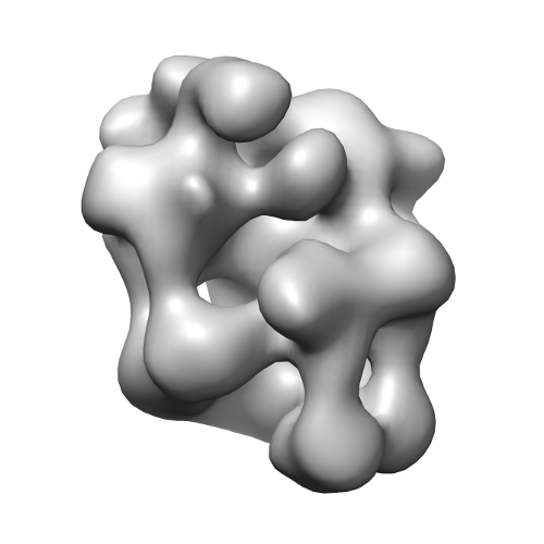 emd_2321.png
 http://ftp.pdbj.org/pub/emdb/structures/EMD-2321


 Sample components
 Processing
 Electron microscopy