 Movie
Movie Controller

[English] 日本語
 Yorodumi
- EMDB-2315: Electron crystallographic studies on IP39 of Euglena gracilis with Fab -
+ Open data

- Basic information
| Entry | Database: EMDB / ID: EMD-2315 | |||||||||
|---|---|---|---|---|---|---|---|---|---|---|
| Title | Electron crystallographic studies on IP39 of Euglena gracilis with Fab | |||||||||
 Map data Map data | 3D reconstruction of 2D crystals of IP39 bound with Fab | |||||||||
 Sample Sample |
| |||||||||
| Function / homology | membrane => GO:0016020 / Alpha-type IP39 Function and homology information Function and homology information | |||||||||
| Biological species |  Euglena gracilis (euglena) Euglena gracilis (euglena) | |||||||||
| Method | electron crystallography / cryo EM / Resolution: 10.0 Å | |||||||||
 Authors Authors | Suzuki H / Ito Y / Yamazaki Y / Mineta K / Uji M / Abe K / Tani K / Fujiyoshi Y / Tsukita S | |||||||||
 Citation Citation | Journal: Nat Commun / Year: 2013 Title: The four-transmembrane protein IP39 of Euglena forms strands by a trimeric unit repeat. Authors: Hiroshi Suzuki / Yasuyuki Ito / Yuji Yamazaki / Katsuhiko Mineta / Masami Uji / Kazuhiro Abe / Kazutoshi Tani / Yoshinori Fujiyoshi / Sachiko Tsukita /  Abstract: Euglenoid flagellates have striped surface structures comprising pellicles, which allow the cell shape to vary from rigid to flexible during the characteristic movement of the flagellates. In Euglena ...Euglenoid flagellates have striped surface structures comprising pellicles, which allow the cell shape to vary from rigid to flexible during the characteristic movement of the flagellates. In Euglena gracilis, the pellicular strip membranes are covered with paracrystalline arrays of a major integral membrane protein, IP39, a putative four-membrane-spanning protein with the conserved sequence motif of the PMP-22/EMP/MP20/Claudin superfamily. Here we report the three-dimensional structure of Euglena IP39 determined by electron crystallography. Two-dimensional crystals of IP39 appear to form a striated pattern of antiparallel double-rows in which trimeric IP39 units are longitudinally polymerised, resulting in continuously extending zigzag-shaped lines. Structural analysis revealed an asymmetric molecular arrangement in the trimer, and suggested that at least four different interactions between neighbouring protomers are involved. A combination of such multiple interactions would be important for linear strand formation of membrane proteins in a lipid bilayer. | |||||||||
| History |
|
- Structure visualization
| Movie |
Movie viewer |
|---|---|
| Structure viewer | EM map: SurfViewMolmilJmol/JSmol |
| Supplemental images |

- Downloads & links
-EMDB archive
| Map data | emd_2315.map.gz | 400.6 KB | EMDB map data format | |
|---|---|---|---|---|
| Header (meta data) | emd-2315-v30.xmlemd-2315.xml | 10.3 KB 10.3 KB | Display Display | EMDB header |
| Images | 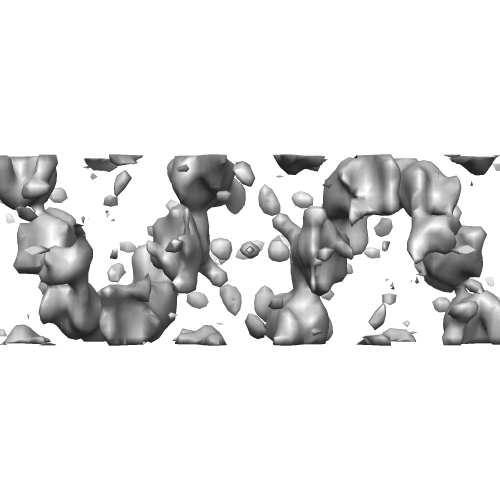 EMD-2315.png EMD-2315.png | 68.8 KB | ||
| Archive directory |  http://ftp.pdbj.org/pub/emdb/structures/EMD-2315ftp://ftp.pdbj.org/pub/emdb/structures/EMD-2315 http://ftp.pdbj.org/pub/emdb/structures/EMD-2315ftp://ftp.pdbj.org/pub/emdb/structures/EMD-2315 | HTTPS FTP |
-Validation report
| Summary document | emd_2315_validation.pdf.gz | 197.2 KB | Display | EMDB validaton report |
|---|---|---|---|---|
| Full document | emd_2315_full_validation.pdf.gz | 196.3 KB | Display | |
| Data in XML | emd_2315_validation.xml.gz | 4.5 KB | Display | |
| Arichive directory | https://ftp.pdbj.org/pub/emdb/validation_reports/EMD-2315ftp://ftp.pdbj.org/pub/emdb/validation_reports/EMD-2315 | HTTPS FTP |
-Related structure data

-Links
| EMDB pages | EMDB (EBI/PDBe) / EMDataResource |
|---|
-Map
| File | Download / File: emd_2315.map.gz / Format: CCP4 / Size: 565.4 KB / Type: IMAGE STORED AS FLOATING POINT NUMBER (4 BYTES) | ||||||||||||||||||||||||||||||||||||||||||||||||||||||||||||||||||||
|---|---|---|---|---|---|---|---|---|---|---|---|---|---|---|---|---|---|---|---|---|---|---|---|---|---|---|---|---|---|---|---|---|---|---|---|---|---|---|---|---|---|---|---|---|---|---|---|---|---|---|---|---|---|---|---|---|---|---|---|---|---|---|---|---|---|---|---|---|---|
| Annotation | 3D reconstruction of 2D crystals of IP39 bound with Fab | ||||||||||||||||||||||||||||||||||||||||||||||||||||||||||||||||||||
| Voxel size | X: 2.42083 Å / Y: 2.47083 Å / Z: 2.5 Å | ||||||||||||||||||||||||||||||||||||||||||||||||||||||||||||||||||||
| Density |
| ||||||||||||||||||||||||||||||||||||||||||||||||||||||||||||||||||||
| Symmetry | Space group: 1 | ||||||||||||||||||||||||||||||||||||||||||||||||||||||||||||||||||||
| Details | EMDB XML:
CCP4 map header:
| ||||||||||||||||||||||||||||||||||||||||||||||||||||||||||||||||||||
-Supplemental data
- Sample components
-Entire : Euglena IP39
| Entire | Name: Euglena IP39 |
|---|---|
| Components |
|
-Supramolecule #1000: Euglena IP39
| Supramolecule | Name: Euglena IP39 / type: sample / ID: 1000 / Details: 2D array / Oligomeric state: Trimer / Number unique components: 12 |
|---|---|
| Molecular weight | Theoretical: 39 KDa |
-Macromolecule #1: Alpha-type IP39
| Macromolecule | Name: Alpha-type IP39 / type: protein_or_peptide / ID: 1 / Name.synonym: Major integral plasma membrane protein / Number of copies: 12 / Oligomeric state: trimer / Recombinant expression: No / Database: NCBI |
|---|---|
| Source (natural) | Organism: Euglena gracilis (euglena) / Location in cell: Plasma membrane |
| Molecular weight | Theoretical: 39 KDa |
| Sequence | UniProtKB: Alpha-type IP39 |
-Experimental details
-Structure determination
| Method | cryo EM |
|---|---|
 Processing Processing | electron crystallography |
| Aggregation state | 2D array |
-Sample preparation
| Concentration | 0.35 mg/mL |
|---|---|
| Buffer | pH: 7 Details: 10 mM Hepes (pH7.0), 150 mM NaCl, 1 mM MgCl2, 30% (v/v) glycerol |
| Grid | Details: carbon sandwich method |
| Vitrification | Cryogen name: NITROGEN / Instrument: LEICA KF80 / Details: Vitrification carried out at 4 degree. |
| Details | Crystal growth by dialysis |
| Crystal formation | Details: Crystal growth by dialysis |
- Electron microscopy
| Microscope | JEOL KYOTO-3000SFF |
|---|---|
| Date | Dec 28, 2011 |
| Image recording | Category: FILM / Film or detector model: KODAK SO-163 FILM / Digitization - Scanner: ZEISS SCAI / Digitization - Sampling interval: 7 µm / Number real images: 198 / Bits/pixel: 8 |
| Tilt angle min | 0 |
| Electron beam | Acceleration voltage: 300 kV / Electron source:  FIELD EMISSION GUN FIELD EMISSION GUN |
| Electron optics | Calibrated magnification: 39500 / Illumination mode: FLOOD BEAM / Imaging mode: BRIGHT FIELD / Cs: 1.6 mm / Nominal defocus max: 2.3 µm / Nominal defocus min: 0.62 µm / Nominal magnification: 40000 |
| Sample stage | Specimen holder: Helium cooled / Specimen holder model: JEOL / Tilt angle max: 45 / Tilt series - Axis1 - Min angle: 0 ° / Tilt series - Axis1 - Max angle: 45 ° |
-Image processing
| Details | MRC package suite |
|---|---|
| Final reconstruction | Resolution.type: BY AUTHOR / Resolution: 10.0 Å / Resolution method: OTHER / Software - Name: MRC |
| Crystal parameters | Unit cell - A: 174.3 Å / Unit cell - B: 59.3 Å / Unit cell - C: 200.0 Å / Unit cell - γ: 90.0 ° / Unit cell - α: 90.0 ° / Unit cell - β: 90.0 ° / Plane group: P 2 |
| CTF correction | Details: Each micrographs |
