[English] 日本語
 Yorodumi
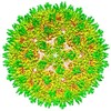
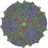
- EMDB-2278: cryoEM structure of hepatitis B virus core assembled from full-le... -
+ Open data
- Basic information
| Entry | Database: EMDB / ID: EMD-2278 | |||||||||
|---|---|---|---|---|---|---|---|---|---|---|
| Title | cryoEM structure of hepatitis B virus core assembled from full-length core protein | |||||||||
 Map data Map data | Reconstruction of HBV | |||||||||
 Sample Sample |
| |||||||||
 Keywords Keywords | hepatitis B virus core antigen (HBc) / HBV capsid maturation and envelopment | |||||||||
| Function / homology |  Function and homology information Function and homology informationvirus-mediated perturbation of host defense response => GO:0019049 / microtubule-dependent intracellular transport of viral material towards nucleus / T=4 icosahedral viral capsid / : / viral penetration into host nucleus / host cell cytoplasm / symbiont entry into host cell / structural molecule activity /  DNA binding / DNA binding /  RNA binding / extracellular region RNA binding / extracellular regionSimilarity search - Function | |||||||||
| Biological species |    Hepatitis B virus Hepatitis B virus | |||||||||
| Method |  single particle reconstruction / single particle reconstruction /  cryo EM / Resolution: 3.5 Å cryo EM / Resolution: 3.5 Å | |||||||||
 Authors Authors | Yu X / Jin L / Jih J / Shih C / Zhou ZH | |||||||||
 Citation Citation |  Journal: PLoS One / Year: 2013 Journal: PLoS One / Year: 2013Title: 3.5Å cryoEM structure of hepatitis B virus core assembled from full-length core protein. Authors: Xuekui Yu / Lei Jin / Jonathan Jih / Chiaho Shih / Z Hong Zhou /  Abstract: The capsid shell of infectious hepatitis B virus (HBV) is composed of 240 copies of a single protein called HBV core antigen (HBc). An atomic model of a core assembled from truncated HBc was ...The capsid shell of infectious hepatitis B virus (HBV) is composed of 240 copies of a single protein called HBV core antigen (HBc). An atomic model of a core assembled from truncated HBc was determined previously by X-ray crystallography. In an attempt to obtain atomic structural information of HBV core in a near native, non-crystalline environment, we reconstructed a 3.5Å-resolution structure of a recombinant core assembled from full-length HBc by cryo electron microscopy (cryoEM) and derived an atomic model. The structure shows that the 240 molecules of full-length HBc form a core with two layers. The outer layer, composed of the N-terminal assembly domain, is similar to the crystal structure of the truncated HBc, but has three differences. First, unlike the crystal structure, our cryoEM structure shows no disulfide bond between the Cys61 residues of the two subunits within the dimer building block, indicating such bond is not required for core formation. Second, our cryoEM structure reveals up to four more residues in the linker region (amino acids 140-149). Third, the loops in the cryoEM structures containing this linker region in subunits B and C are oriented differently (~30° and ~90°) from their counterparts in the crystal structure. The inner layer, composed of the C-terminal arginine-rich domain (ARD) and the ARD-bound RNAs, is partially-ordered and connected with the outer layer through linkers positioned around the two-fold axes. Weak densities emanate from the rims of positively charged channels through the icosahedral three-fold and local three-fold axes. We attribute these densities to the exposed portions of some ARDs, thus explaining ARD's accessibility by proteases and antibodies. Our data supports a role of ARD in mediating communication between inside and outside of the core during HBV maturation and envelopment. | |||||||||
| History |
|
- Structure visualization
| Movie |
 Movie viewer Movie viewer |
|---|---|
| Structure viewer | EM map:  SurfView SurfView Molmil Molmil Jmol/JSmol Jmol/JSmol |
| Supplemental images |
- Downloads & links
-EMDB archive
| Map data |  emd_2278.map.gz emd_2278.map.gz | 180.1 MB |  EMDB map data format EMDB map data format | |
|---|---|---|---|---|
| Header (meta data) |  emd-2278-v30.xml emd-2278-v30.xml emd-2278.xml emd-2278.xml | 8.3 KB 8.3 KB | Display Display |  EMDB header EMDB header |
| Images |  EMD-2278-HBV.jpg EMD-2278-HBV.jpg | 134.1 KB | ||
| Archive directory |  http://ftp.pdbj.org/pub/emdb/structures/EMD-2278 http://ftp.pdbj.org/pub/emdb/structures/EMD-2278 ftp://ftp.pdbj.org/pub/emdb/structures/EMD-2278 ftp://ftp.pdbj.org/pub/emdb/structures/EMD-2278 | HTTPS FTP |
-Related structure data
| Related structure data |  3j2vMC M: atomic model generated by this map C: citing same article ( |
|---|---|
| Similar structure data |
- Links
Links
| EMDB pages |  EMDB (EBI/PDBe) / EMDB (EBI/PDBe) /  EMDataResource EMDataResource |
|---|---|
| Related items in Molecule of the Month |
- Map
Map
| File |  Download / File: emd_2278.map.gz / Format: CCP4 / Size: 317.3 MB / Type: IMAGE STORED AS FLOATING POINT NUMBER (4 BYTES) Download / File: emd_2278.map.gz / Format: CCP4 / Size: 317.3 MB / Type: IMAGE STORED AS FLOATING POINT NUMBER (4 BYTES) | ||||||||||||||||||||||||||||||||||||||||||||||||||||||||||||||||||||
|---|---|---|---|---|---|---|---|---|---|---|---|---|---|---|---|---|---|---|---|---|---|---|---|---|---|---|---|---|---|---|---|---|---|---|---|---|---|---|---|---|---|---|---|---|---|---|---|---|---|---|---|---|---|---|---|---|---|---|---|---|---|---|---|---|---|---|---|---|---|
| Annotation | Reconstruction of HBV | ||||||||||||||||||||||||||||||||||||||||||||||||||||||||||||||||||||
| Voxel size | X=Y=Z: 0.9333 Å | ||||||||||||||||||||||||||||||||||||||||||||||||||||||||||||||||||||
| Density |
| ||||||||||||||||||||||||||||||||||||||||||||||||||||||||||||||||||||
| Symmetry | Space group: 1 | ||||||||||||||||||||||||||||||||||||||||||||||||||||||||||||||||||||
| Details | EMDB XML:
CCP4 map header:
| ||||||||||||||||||||||||||||||||||||||||||||||||||||||||||||||||||||
-Supplemental data
- Sample components
-Entire : Hepatitis B virus core assembled from full-length core protein
| Entire | Name: Hepatitis B virus core assembled from full-length core protein |
|---|---|
| Components |
|
-Supramolecule #1000: Hepatitis B virus core assembled from full-length core protein
| Supramolecule | Name: Hepatitis B virus core assembled from full-length core protein type: sample / ID: 1000 / Oligomeric state: Icosahedral particle / Number unique components: 1 |
|---|---|
| Molecular weight | Experimental: 5 MDa / Theoretical: 5 MDa / Method: Based on amino acid sequences |
-Supramolecule #1: Hepatitis B virus
| Supramolecule | Name: Hepatitis B virus / type: virus / ID: 1 / NCBI-ID: 10407 / Sci species name: Hepatitis B virus / Virus type: VIRION / Virus isolate: SPECIES / Virus enveloped: No / Virus empty: No |
|---|---|
| Host (natural) | Organism:   Homo sapiens (human) / synonym: VERTEBRATES Homo sapiens (human) / synonym: VERTEBRATES |
| Host system | Organism:   Escherichia coli (E. coli) / Recombinant plasmid: pET-11a Escherichia coli (E. coli) / Recombinant plasmid: pET-11a |
| Molecular weight | Experimental: 5 MDa / Theoretical: 5 MDa |
| Virus shell | Shell ID: 1 / Diameter: 360 Å / T number (triangulation number): 4 |
-Experimental details
-Structure determination
| Method |  cryo EM cryo EM |
|---|---|
 Processing Processing |  single particle reconstruction single particle reconstruction |
| Aggregation state | particle |
- Sample preparation
| Vitrification | Cryogen name: NITROGEN / Chamber humidity: 100 % / Chamber temperature: 120 K / Instrument: FEI VITROBOT MARK II |
|---|
- Electron microscopy
| Microscope | FEI TITAN KRIOS |
|---|---|
| Electron beam | Acceleration voltage: 300 kV / Electron source:  FIELD EMISSION GUN FIELD EMISSION GUN |
| Electron optics | Illumination mode: FLOOD BEAM / Imaging mode: BRIGHT FIELD Bright-field microscopy / Cs: 2.75 mm / Nominal magnification: 75000 Bright-field microscopy / Cs: 2.75 mm / Nominal magnification: 75000 |
| Sample stage | Specimen holder model: GATAN LIQUID NITROGEN |
| Date | Jan 1, 2009 |
| Image recording | Digitization - Scanner: NIKON SUPER COOLSCAN 9000 / Average electron dose: 25 e/Å2 |
| Experimental equipment |  Model: Titan Krios / Image courtesy: FEI Company |
- Image processing
| CTF correction | Details: Each particle |
|---|---|
| Final reconstruction | Applied symmetry - Point group: I (icosahedral ) / Algorithm: OTHER / Resolution.type: BY AUTHOR / Resolution: 3.5 Å / Resolution method: FSC 0.143 CUT-OFF / Software - Name: IMIRS / Number images used: 8093 ) / Algorithm: OTHER / Resolution.type: BY AUTHOR / Resolution: 3.5 Å / Resolution method: FSC 0.143 CUT-OFF / Software - Name: IMIRS / Number images used: 8093 |
 Movie
Movie Controller


