 Movie
Movie Controller


+ Open data

- Basic information
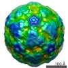
| Entry | Database: EMDB / ID: EMD-2242 | |||||||||
|---|---|---|---|---|---|---|---|---|---|---|
| Title | The Cryo-EM structure of Arabis mosaic virus | |||||||||
 Map data Map data | 3D cryo-EM map of ArMV | |||||||||
 Sample Sample |
| |||||||||
 Keywords Keywords |  Arabis mosaic virus / Nepovirus / cryo-electron microscopy / image processing / molecular dynamics flexible fitting / nematode transmission Arabis mosaic virus / Nepovirus / cryo-electron microscopy / image processing / molecular dynamics flexible fitting / nematode transmission | |||||||||
| Biological species |  Arabis mosaic virus Arabis mosaic virus | |||||||||
| Method | single particle reconstruction / cryo EM / Resolution: 6.5 Å | |||||||||
 Authors Authors | Lai-Kee-Him J / Schellenberger P / Dumas C / Richard E / Trapani S / Komar V / Demangeat G / Ritzenthaler C / Bron P | |||||||||
 Citation Citation | Journal: J Struct Biol / Year: 2013 Title: The backbone model of the Arabis mosaic virus reveals new insights into functional domains of Nepovirus capsid. Authors: Joséphine Lai-Kee-Him / Pascale Schellenberger / Christian Dumas / Eric Richard / Stefano Trapani / Véronique Komar / Gerard Demangeat / Christophe Ritzenthaler / Patrick Bron /  Abstract: Arabis mosaic virus (ArMV) and Grapevine fanleaf virus (GFLV) are two picorna-like viruses from the genus Nepovirus, consisting in a bipartite RNA genome encapsidated into a 30 nm icosahedral viral ...Arabis mosaic virus (ArMV) and Grapevine fanleaf virus (GFLV) are two picorna-like viruses from the genus Nepovirus, consisting in a bipartite RNA genome encapsidated into a 30 nm icosahedral viral particle formed by 60 copies of a single capsid protein (CP). They are responsible for a severe degeneration of grapevines that occurs in most vineyards worldwide. Although sharing a high level of sequence identity between their CP, ArMV is transmitted exclusively by the ectoparasitic nematode Xiphinema diversicaudatum whereas GFLV is specifically transmitted by the nematode X. index. The structural determinants involved in the transmission specificity of both viruses map solely to their respective CP. Recently, reverse genetic and crystallographic studies on GFLV revealed that a positively charged pocket in the CP B domain located at the virus surface may be responsible for vector specificity. To go further into delineating the coat protein determinants involved in transmission specificity, we determined the 6.5 Å resolution cryo-electron microscopy structure of ArMV and used homology modeling and flexible fitting approaches to build its pseudo-atomic structure. This study allowed us to resolve ArMV CP architecture and delineate connections between ArMV capsid shell and its RNA. Comparison of ArMV and GFLV CPs reveals structural differences in the B domain pocket, thus strengthening the hypothesis of a key role of this region in the viral transmission specificity and identifies new potential functional domains of Nepovirus capsid. | |||||||||
| History |
|
- Structure visualization
| Movie |
 Movie viewer Movie viewer |
|---|---|
| Structure viewer | EM map: SurfViewMolmilJmol/JSmol |
| Supplemental images |

- Downloads & links
-EMDB archive
| Map data | emd_2242.map.gz | 31.1 MB | EMDB map data format | |
|---|---|---|---|---|
| Header (meta data) | emd-2242-v30.xmlemd-2242.xml | 9.9 KB 9.9 KB | Display Display | EMDB header |
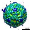
| Images | 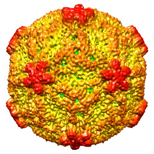 image2242mattsversion.png image2242mattsversion.png | 339.5 KB | ||
| Archive directory |  http://ftp.pdbj.org/pub/emdb/structures/EMD-2242ftp://ftp.pdbj.org/pub/emdb/structures/EMD-2242 http://ftp.pdbj.org/pub/emdb/structures/EMD-2242ftp://ftp.pdbj.org/pub/emdb/structures/EMD-2242 | HTTPS FTP |
-Related structure data
| Similar structure data |
|---|

-Links
| EMDB pages | EMDB (EBI/PDBe) / EMDataResource |
|---|
-Map
| File | Download / File: emd_2242.map.gz / Format: CCP4 / Size: 82.7 MB / Type: IMAGE STORED AS FLOATING POINT NUMBER (4 BYTES) | ||||||||||||||||||||||||||||||||||||||||||||||||||||||||||||||||||||
|---|---|---|---|---|---|---|---|---|---|---|---|---|---|---|---|---|---|---|---|---|---|---|---|---|---|---|---|---|---|---|---|---|---|---|---|---|---|---|---|---|---|---|---|---|---|---|---|---|---|---|---|---|---|---|---|---|---|---|---|---|---|---|---|---|---|---|---|---|---|
| Annotation | 3D cryo-EM map of ArMV | ||||||||||||||||||||||||||||||||||||||||||||||||||||||||||||||||||||
| Voxel size | X=Y=Z: 1.49 Å | ||||||||||||||||||||||||||||||||||||||||||||||||||||||||||||||||||||
| Density |
| ||||||||||||||||||||||||||||||||||||||||||||||||||||||||||||||||||||
| Symmetry | Space group: 1 | ||||||||||||||||||||||||||||||||||||||||||||||||||||||||||||||||||||
| Details | EMDB XML:
CCP4 map header:
| ||||||||||||||||||||||||||||||||||||||||||||||||||||||||||||||||||||
-Supplemental data
- Sample components
-Entire : Purified ArMV particle containing its RNA
| Entire | Name: Purified ArMV particle containing its RNA |
|---|---|
| Components |
|
-Supramolecule #1000: Purified ArMV particle containing its RNA
| Supramolecule | Name: Purified ArMV particle containing its RNA / type: sample / ID: 1000 / Number unique components: 2 |
|---|
-Supramolecule #1: Arabis mosaic virus
| Supramolecule | Name: Arabis mosaic virus / type: virus / ID: 1 / Name.synonym: ArMV Details: The virion is purified from infected plants and contains its RNA NCBI-ID: 12271 / Sci species name: Arabis mosaic virus / Virus type: VIRION / Virus isolate: SPECIES / Virus enveloped: No / Virus empty: No / Syn species name: ArMV |
|---|---|
| Host (natural) | Organism:  Chenopodium quinoa (quinoa) / synonym: PLANTAE(HIGHER PLANTS) Chenopodium quinoa (quinoa) / synonym: PLANTAE(HIGHER PLANTS) |
| Virus shell | Shell ID: 1 / Diameter: 300 Å / T number (triangulation number): 3 |
-Experimental details
-Structure determination
| Method | cryo EM |
|---|---|
 Processing Processing | single particle reconstruction |
| Aggregation state | particle |
-Sample preparation
| Concentration | 2 mg/mL |
|---|---|
| Buffer | pH: 7 Details: 15 mM sodium phosphate and 5 mM potassium phosphate pH 7.0 |
| Grid | Details: Quantifoil R 2/2 grids (Quantifoil Micro Tools GmbH, Jena, Germany) |
| Vitrification | Cryogen name: ETHANE / Chamber humidity: 98 % / Chamber temperature: 103.15 K / Instrument: GATAN CRYOPLUNGE 3 Method: blotted for 1s and then flash frozen in liquid ethane |
- Electron microscopy
| Microscope | JEOL 2200FS |
|---|---|
| Electron beam | Acceleration voltage: 200 kV / Electron source: FIELD EMISSION GUN |
| Electron optics | Calibrated magnification: 46980 / Illumination mode: FLOOD BEAM / Imaging mode: BRIGHT FIELDBright-field microscopy / Cs: 2 mm / Nominal defocus max: 2.5 µm / Nominal defocus min: 1.2 µm / Nominal magnification: 50000 |
| Specialist optics | Energy filter - Name: omega / Energy filter - Lower energy threshold: 0.0 eV / Energy filter - Upper energy threshold: 20.0 eV |
| Sample stage | Specimen holder: gatan 626 / Specimen holder model: GATAN LIQUID NITROGEN |
| Temperature | Average: 93.15 K |
| Alignment procedure | Legacy - Astigmatism: Objective lens astigmatism was corrected at 200,000 times magnification |
| Date | Sep 1, 2011 |
| Image recording | Category: FILM / Film or detector model: KODAK SO-163 FILM / Digitization - Scanner: NIKON SUPER COOLSCAN 9000 / Digitization - Sampling interval: 7 µm / Number real images: 400 / Average electron dose: 18 e/Å2 / Bits/pixel: 8 |
| Tilt angle min | 0 |
| Tilt angle max | 0 |
-Image processing
| CTF correction | Details: Each particle |
|---|---|
| Final reconstruction | Applied symmetry - Point group: I (icosahedral) / Resolution.type: BY AUTHOR / Resolution: 6.5 Å / Resolution method: FSC 0.5 CUT-OFF / Software - Name: BOXER, CTFFIND3, CTFMIX, AUTO3DEM / Number images used: 7009 |
| Details | Image processing was performed using AUTO3DEM package |
