 Movie
Movie Controller


+ Open data

- Basic information
| Entry | Database: EMDB / ID: EMD-2220 | |||||||||
|---|---|---|---|---|---|---|---|---|---|---|
| Title | Cryo-EM structure of gastric H+,K+-ATPase with bound K+ | |||||||||
 Map data Map data | Reconstruction of H,K-ATPase with bound K | |||||||||
 Sample Sample |
| |||||||||
 Keywords Keywords |  P-type ATPase / proton pump P-type ATPase / proton pump | |||||||||
| Function / homology | ATP biosynthetic process / P-type ATPase, A domain superfamily Function and homology information Function and homology information | |||||||||
| Biological species |  Sus scrofa (pig) Sus scrofa (pig) | |||||||||
| Method | electron crystallography / cryo EM / Resolution: 8.0 Å | |||||||||
 Authors Authors | Abe K / Tani K / Friedrich T / Fujiyoshi Y | |||||||||
 Citation Citation | Journal: Proc Natl Acad Sci U S A / Year: 2012 Title: Cryo-EM structure of gastric H+,K+-ATPase with a single occupied cation-binding site. Authors: Kazuhiro Abe / Kazutoshi Tani / Thomas Friedrich / Yoshinori Fujiyoshi /  Abstract: Gastric H(+),K(+)-ATPase is responsible for gastric acid secretion. ATP-driven H(+) uptake into the stomach is efficiently accomplished by the exchange of an equal amount of K(+), resulting in a ...Gastric H(+),K(+)-ATPase is responsible for gastric acid secretion. ATP-driven H(+) uptake into the stomach is efficiently accomplished by the exchange of an equal amount of K(+), resulting in a luminal pH close to 1. Because of the limited free energy available for ATP hydrolysis, the stoichiometry of transported cations is thought to vary from 2H(+)/2K(+) to 1H(+)/1K(+) per hydrolysis of one ATP molecule as the luminal pH decreases, although direct evidence for this hypothesis has remained elusive. Here, we show, using the phosphate analog aluminum fluoride (AlF) and a K(+) congener (Rb(+)), the 8-Å resolution structure of H(+),K(+)-ATPase in the transition state of dephosphorylation, (Rb(+))E2~AlF, which is distinct from the preceding Rb(+)-free E2P state. A strong density located in the transmembrane cation-binding site of (Rb(+))E2~AlF highly likely represents a single bound Rb(+) ion, which is clearly different from the Rb(+)-free E2AlF or K(+)-bound (K(+))E2~AlF structures. Measurement of radioactive (86)Rb(+) binding suggests that the binding stoichiometry varies depending on the pH, and approximately half of the amount of Rb(+) is bound under acidic crystallization conditions compared with at a neutral pH. These data represent structural and biochemical evidence for the 1H(+)/1K(+)/1ATP transport mode of H(+),K(+)-ATPase, which is a prerequisite for generation of the 10(6)-fold proton gradient in terms of thermodynamics. Together with the released E2P-stabilizing interaction between the β subunit's N terminus and the P domain observed in the (Rb(+))E2~AlF structure, we propose a refined vectorial transport model of H(+),K(+)-ATPase, which must prevail against the highly acidic state of the gastric lumen. | |||||||||
| History |
|
- Structure visualization
| Movie |
Movie viewer |
|---|---|
| Structure viewer | EM map: SurfViewMolmilJmol/JSmol |
| Supplemental images |

- Downloads & links
-EMDB archive
| Map data | emd_2220.map.gz | 2 MB | EMDB map data format | |
|---|---|---|---|---|
| Header (meta data) | emd-2220-v30.xmlemd-2220.xml | 10.1 KB 10.1 KB | Display Display | EMDB header |
| Images | 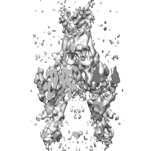 EMD-2220.jpg EMD-2220.jpg | 49.6 KB | ||
| Archive directory |  http://ftp.pdbj.org/pub/emdb/structures/EMD-2220ftp://ftp.pdbj.org/pub/emdb/structures/EMD-2220 http://ftp.pdbj.org/pub/emdb/structures/EMD-2220ftp://ftp.pdbj.org/pub/emdb/structures/EMD-2220 | HTTPS FTP |
-Related structure data

-Links
| EMDB pages | EMDB (EBI/PDBe) / EMDataResource |
|---|
-Map
| File | Download / File: emd_2220.map.gz / Format: CCP4 / Size: 2.7 MB / Type: IMAGE STORED AS FLOATING POINT NUMBER (4 BYTES) | ||||||||||||||||||||||||||||||||||||||||||||||||||||||||||||||||||||
|---|---|---|---|---|---|---|---|---|---|---|---|---|---|---|---|---|---|---|---|---|---|---|---|---|---|---|---|---|---|---|---|---|---|---|---|---|---|---|---|---|---|---|---|---|---|---|---|---|---|---|---|---|---|---|---|---|---|---|---|---|---|---|---|---|---|---|---|---|---|
| Annotation | Reconstruction of H,K-ATPase with bound K | ||||||||||||||||||||||||||||||||||||||||||||||||||||||||||||||||||||
| Voxel size | X: 1.8433 Å / Y: 1.9583 Å / Z: 2 Å | ||||||||||||||||||||||||||||||||||||||||||||||||||||||||||||||||||||
| Density |
| ||||||||||||||||||||||||||||||||||||||||||||||||||||||||||||||||||||
| Symmetry | Space group: 1 | ||||||||||||||||||||||||||||||||||||||||||||||||||||||||||||||||||||
| Details | EMDB XML:
CCP4 map header:
| ||||||||||||||||||||||||||||||||||||||||||||||||||||||||||||||||||||
-Supplemental data
- Sample components
-Entire : Pig Gastric H,K-ATPase
| Entire | Name: Pig Gastric H,K-ATPase |
|---|---|
| Components |
|
-Supramolecule #1000: Pig Gastric H,K-ATPase
| Supramolecule | Name: Pig Gastric H,K-ATPase / type: sample / ID: 1000 / Oligomeric state: One alpha and one beta chain of HK-ATPase / Number unique components: 1 |
|---|---|
| Molecular weight | Theoretical: 150 KDa |
-Macromolecule #1: POTASSIUM-TRANSPORTING ATPASE
| Macromolecule | Name: POTASSIUM-TRANSPORTING ATPASE / type: protein_or_peptide / ID: 1 / Name.synonym: H+,K+-ATPase / Number of copies: 2 / Oligomeric state: Dimer / Recombinant expression: No / Database: NCBI |
|---|---|
| Source (natural) | Organism: Sus scrofa (pig) / synonym: Pig / Tissue: stomach / Location in cell: Plasma membrane |
| Molecular weight | Theoretical: 150 KDa |
| Sequence | GO: ATP biosynthetic process / InterPro: P-type ATPase, A domain superfamily |
-Experimental details
-Structure determination
| Method | cryo EM |
|---|---|
 Processing Processing | electron crystallography |
| Aggregation state | 2D array |
-Sample preparation
| Concentration | 8 mg/mL |
|---|---|
| Buffer | pH: 4.8 Details: 20mM propionate, 1mM ADP, 3mM DTT, ph4.8-4.9 adjusted by Tris, 1mM MgCl2, 1mM AlCl3, 4mM NaF, 10mM KCl |
| Grid | Details: Molybdenum grid |
| Vitrification | Cryogen name: NITROGEN / Instrument: LEICA KF80 Details: The grids were blotted with filter paper and fast frozen into liquid nitrogen |
| Details | Crystals grown by dialysis |
| Crystal formation | Details: Crystals grown by dialysis |
- Electron microscopy
| Microscope | JEOL KYOTO-3000SFF |
|---|---|
| Electron beam | Acceleration voltage: 300 kV / Electron source: FIELD EMISSION GUN |
| Electron optics | Calibrated magnification: 39500 / Illumination mode: FLOOD BEAM / Imaging mode: BRIGHT FIELDBright-field microscopy / Cs: 1.6 mm / Nominal defocus max: 3.88 µm / Nominal defocus min: 0.67 µm / Nominal magnification: 40000 |
| Sample stage | Specimen holder: Helium cooled / Specimen holder model: JEOL / Tilt angle min: -60 / Tilt angle max: 60 / Tilt series - Axis1 - Min angle: -60 ° / Tilt series - Axis1 - Max angle: 60 ° |
| Date | Feb 15, 2012 |
| Image recording | Category: FILM / Film or detector model: KODAK SO-163 FILM / Digitization - Scanner: ZEISS SCAI / Digitization - Sampling interval: 7 µm / Number real images: 278 / Bits/pixel: 12 |
-Image processing
| Crystal parameters | Unit cell - A: 141.0 Å / Unit cell - B: 110.6 Å / Unit cell - C: 320.0 Å / Unit cell - γ: 90.0 ° / Unit cell - α: 90.0 ° / Unit cell - β: 90.0 ° / Plane group: P 2 21 21 |
|---|---|
| CTF correction | Details: Each image |
| Final reconstruction | Resolution.type: BY AUTHOR / Resolution: 8.0 Å / Software - Name: MRC |
| Details | Images were processed using MRC suite. |
