Journal: Proc Natl Acad Sci U S A / Year: 2012 Title: Cryo-EM structure of gastric H+,K+-ATPase with a single occupied cation-binding site. Authors: Kazuhiro Abe / Kazutoshi Tani / Thomas Friedrich / Yoshinori Fujiyoshi / Abstract: Gastric H(+),K(+)-ATPase is responsible for gastric acid secretion. ATP-driven H(+) uptake into the stomach is efficiently accomplished by the exchange of an equal amount of K(+), resulting in a ...Gastric H(+),K(+)-ATPase is responsible for gastric acid secretion. ATP-driven H(+) uptake into the stomach is efficiently accomplished by the exchange of an equal amount of K(+), resulting in a luminal pH close to 1. Because of the limited free energy available for ATP hydrolysis, the stoichiometry of transported cations is thought to vary from 2H(+)/2K(+) to 1H(+)/1K(+) per hydrolysis of one ATP molecule as the luminal pH decreases, although direct evidence for this hypothesis has remained elusive. Here, we show, using the phosphate analog aluminum fluoride (AlF) and a K(+) congener (Rb(+)), the 8-Å resolution structure of H(+),K(+)-ATPase in the transition state of dephosphorylation, (Rb(+))E2~AlF, which is distinct from the preceding Rb(+)-free E2P state. A strong density located in the transmembrane cation-binding site of (Rb(+))E2~AlF highly likely represents a single bound Rb(+) ion, which is clearly different from the Rb(+)-free E2AlF or K(+)-bound (K(+))E2~AlF structures. Measurement of radioactive (86)Rb(+) binding suggests that the binding stoichiometry varies depending on the pH, and approximately half of the amount of Rb(+) is bound under acidic crystallization conditions compared with at a neutral pH. These data represent structural and biochemical evidence for the 1H(+)/1K(+)/1ATP transport mode of H(+),K(+)-ATPase, which is a prerequisite for generation of the 10(6)-fold proton gradient in terms of thermodynamics. Together with the released E2P-stabilizing interaction between the β subunit's N terminus and the P domain observed in the (Rb(+))E2~AlF structure, we propose a refined vectorial transport model of H(+),K(+)-ATPase, which must prevail against the highly acidic state of the gastric lumen.
History
Deposition
Oct 12, 2012
-
Header (metadata) release
Nov 14, 2012
-
Map release
Nov 14, 2012
-
Update
Nov 14, 2012
-
Current status
Nov 14, 2012
Processing site: PDBe / Status: Released
-
Structure visualization
Movie
Surface view with section colored by density value
Download / File: emd_2219.map.gz / Format: CCP4 / Size: 2.7 MB / Type: IMAGE STORED AS FLOATING POINT NUMBER (4 BYTES)
Annotation
Reconstruction of H+,K+-ATPase with Rb+
Voxel size
X: 1.8433 Å / Y: 1.9583 Å / Z: 2 Å
Density
Contour Level
By AUTHOR: 1.2 / Movie #1: 1.2
Minimum - Maximum
-4.47049999 - 6.63910007
Average (Standard dev.)
-0.00531801 (±0.99277723)
Symmetry
Space group: 1
Details
EMDB XML:
Map geometry
Axis order
Y
X
Z
Origin
-36
-30
-80
Dimensions
73
61
161
Spacing
61
73
161
Cell
A: 134.5609 Å / B: 119.4563 Å / C: 322.0 Å α=β=γ: 90.0 °
CCP4 map header:
mode
Image stored as Reals
Å/pix. X/Y/Z
1.843301369863
1.9582950819672
2
M x/y/z
73
61
161
origin x/y/z
0.000
0.000
0.000
length x/y/z
134.561
119.456
322.000
α/β/γ
90.000
90.000
90.000
start NX/NY/NZ
-36
-30
-80
NX/NY/NZ
73
61
161
MAP C/R/S
2
1
3
start NC/NR/NS
-30
-36
-80
NC/NR/NS
61
73
161
D min/max/mean
-4.470
6.639
-0.005
-
Supplemental data
-
Sample components
-
Entire : H+,K+-ATPase bound with Rb+
Entire
Name: H+,K+-ATPase bound with Rb+
Components
Sample: H+,K+-ATPase bound with Rb+
Protein or peptide: POTASSIUM-TRANSPORTING ATPASE
-
Supramolecule #1000: H+,K+-ATPase bound with Rb+
Supramolecule
Name: H+,K+-ATPase bound with Rb+ / type: sample / ID: 1000 / Oligomeric state: One alpha and one beta chain of HK-ATPase / Number unique components: 1
Molecular weight
Theoretical: 150 KDa
-
Macromolecule #1: POTASSIUM-TRANSPORTING ATPASE
Macromolecule
Name: POTASSIUM-TRANSPORTING ATPASE / type: protein_or_peptide / ID: 1 / Number of copies: 2 / Oligomeric state: Dimer / Recombinant expression: No / Database: NCBI
Source (natural)
Organism: Sus scrofa (pig) / synonym: Pig / Tissue: Stomach / Location in cell: Plasma membrane
Molecular weight
Theoretical: 150 KDa
Sequence
GO: ATP biosynthetic process / InterPro: P-type ATPase, A domain superfamily
Specimen holder: Helium cooled / Specimen holder model: JEOL / Tilt angle min: -60 / Tilt angle max: 60 / Tilt series - Axis1 - Min angle: -60 ° / Tilt series - Axis1 - Max angle: 60 °
Date
Mar 23, 2010
Image recording
Category: FILM / Film or detector model: KODAK SO-163 FILM / Digitization - Scanner: ZEISS SCAI / Digitization - Sampling interval: 7 µm / Number real images: 248 / Bits/pixel: 12
-
Image processing
Crystal parameters
Unit cell - A: 141.0 Å / Unit cell - B: 110.6 Å / Unit cell - C: 320.0 Å / Unit cell - γ: 90.0 ° / Unit cell - α: 90.0 ° / Unit cell - β: 90.0 ° / Plane group: P 2 21 21
CTF correction
Details: Each image
Final reconstruction
Resolution.type: BY AUTHOR / Resolution: 8.0 Å / Software - Name: MRC
Details
Images were processed using MRC suite.
+
About Yorodumi
-
News
-
Feb 9, 2022. New format data for meta-information of EMDB entries
New format data for meta-information of EMDB entries
Version 3 of the EMDB header file is now the official format.
The previous official version 1.9 will be removed from the archive.
In the structure databanks used in Yorodumi, some data are registered as the other names, "COVID-19 virus" and "2019-nCoV". Here are the details of the virus and the list of structure data.
Jan 31, 2019. EMDB accession codes are about to change! (news from PDBe EMDB page)
EMDB accession codes are about to change! (news from PDBe EMDB page)
The allocation of 4 digits for EMDB accession codes will soon come to an end. Whilst these codes will remain in use, new EMDB accession codes will include an additional digit and will expand incrementally as the available range of codes is exhausted. The current 4-digit format prefixed with “EMD-” (i.e. EMD-XXXX) will advance to a 5-digit format (i.e. EMD-XXXXX), and so on. It is currently estimated that the 4-digit codes will be depleted around Spring 2019, at which point the 5-digit format will come into force.
The EM Navigator/Yorodumi systems omit the EMD- prefix.
Related info.:Q: What is EMD? / ID/Accession-code notation in Yorodumi/EM Navigator
Yorodumi is a browser for structure data from EMDB, PDB, SASBDB, etc.
This page is also the successor to EM Navigator detail page, and also detail information page/front-end page for Omokage search.
The word "yorodu" (or yorozu) is an old Japanese word meaning "ten thousand". "mi" (miru) is to see.
Related info.:EMDB / PDB / SASBDB / Comparison of 3 databanks / Yorodumi Search / Aug 31, 2016. New EM Navigator & Yorodumi / Yorodumi Papers / Jmol/JSmol / Function and homology information / Changes in new EM Navigator and Yorodumi
 Movie
Movie Controller


 Open data
 Basic information
 Map data
 Sample
 Keywords
 P-type ATPase / Gastric H+/K+-ATPase /
  Function and homology information
 Authors
 Citation
 Structure visualization
 Downloads & links
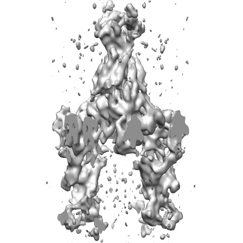 EMD-2219.jpg
 http://ftp.pdbj.org/pub/emdb/structures/EMD-2219


 Sample components
 Processing
 Electron microscopy