 Movie
Movie Controller


+ Open data

- Basic information
| Entry | Database: EMDB / ID: EMD-2171 | |||||||||
|---|---|---|---|---|---|---|---|---|---|---|
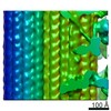
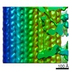
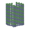
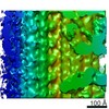
| Title | Staphylothermus marinus 50S ribosomal subunit | |||||||||
 Map data Map data | Staphylothermus marinus 50S ribosomal subunit | |||||||||
 Sample Sample |
| |||||||||
 Keywords Keywords |  archaea / ribosome 50S protein / RNA / kink-turn / protein synthesis / mass spectrometry archaea / ribosome 50S protein / RNA / kink-turn / protein synthesis / mass spectrometry | |||||||||
| Biological species |  Staphylothermus marinus (archaea) Staphylothermus marinus (archaea) | |||||||||
| Method | single particle reconstruction / cryo EM / negative staining / Resolution: 24.0 Å | |||||||||
 Authors Authors | Armache J-P / Anger AM / Marquez V / Frankenberg S / Froehlich T / Villa E / Berninghausen O / Thomm M / Arnold GJ / Beckmann R / Wilson DN | |||||||||
 Citation Citation | Journal: Nucleic Acids Res / Year: 2013 Title: Promiscuous behaviour of archaeal ribosomal proteins: implications for eukaryotic ribosome evolution. Authors: Jean-Paul Armache / Andreas M Anger / Viter Márquez / Sibylle Franckenberg / Thomas Fröhlich / Elizabeth Villa / Otto Berninghausen / Michael Thomm / Georg J Arnold / Roland Beckmann / Daniel N Wilson /  Abstract: In all living cells, protein synthesis occurs on ribonucleoprotein particles called ribosomes. Molecular models have been reported for complete bacterial 70S and eukaryotic 80S ribosomes; however, ...In all living cells, protein synthesis occurs on ribonucleoprotein particles called ribosomes. Molecular models have been reported for complete bacterial 70S and eukaryotic 80S ribosomes; however, only molecular models of large 50S subunits have been reported for archaea. Here, we present a complete molecular model for the Pyrococcus furiosus 70S ribosome based on a 6.6 Å cryo-electron microscopy map. Moreover, we have determined cryo-electron microscopy reconstructions of the Euryarchaeota Methanococcus igneus and Thermococcus kodakaraensis 70S ribosomes and Crenarchaeota Staphylothermus marinus 50S subunit. Examination of these structures reveals a surprising promiscuous behavior of archaeal ribosomal proteins: We observe intersubunit promiscuity of S24e and L8e (L7ae), the latter binding to the head of the small subunit, analogous to S12e in eukaryotes. Moreover, L8e and L14e exhibit intrasubunit promiscuity, being present in two copies per archaeal 50S subunit, with the additional binding site of L14e analogous to the related eukaryotic r-protein L27e. Collectively, these findings suggest insights into the evolution of eukaryotic ribosomal proteins through increased copy number and binding site promiscuity. | |||||||||
| History |
|
- Structure visualization
| Movie |
 Movie viewer Movie viewer |
|---|---|
| Structure viewer | EM map: SurfViewMolmilJmol/JSmol |
| Supplemental images |

- Downloads & links
-EMDB archive
| Map data | emd_2171.map.gz | 10.3 MB | EMDB map data format | |
|---|---|---|---|---|
| Header (meta data) | emd-2171-v30.xmlemd-2171.xml | 9.5 KB 9.5 KB | Display Display | EMDB header |
| Images | 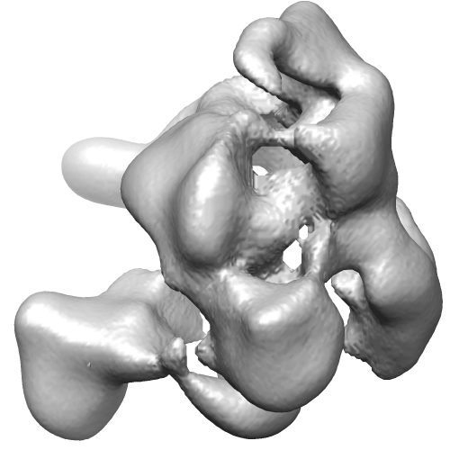 EMD-2171.png EMD-2171.png | 109.8 KB | ||
| Archive directory |  http://ftp.pdbj.org/pub/emdb/structures/EMD-2171ftp://ftp.pdbj.org/pub/emdb/structures/EMD-2171 http://ftp.pdbj.org/pub/emdb/structures/EMD-2171ftp://ftp.pdbj.org/pub/emdb/structures/EMD-2171 | HTTPS FTP |
-Related structure data
-Links
| EMDB pages | EMDB (EBI/PDBe) / EMDataResource |
|---|---|
| Related items in Molecule of the Month |

-Map
| File | Download / File: emd_2171.map.gz / Format: CCP4 / Size: 185.7 MB / Type: IMAGE STORED AS FLOATING POINT NUMBER (4 BYTES) | ||||||||||||||||||||||||||||||||||||||||||||||||||||||||||||||||||||
|---|---|---|---|---|---|---|---|---|---|---|---|---|---|---|---|---|---|---|---|---|---|---|---|---|---|---|---|---|---|---|---|---|---|---|---|---|---|---|---|---|---|---|---|---|---|---|---|---|---|---|---|---|---|---|---|---|---|---|---|---|---|---|---|---|---|---|---|---|---|
| Annotation | Staphylothermus marinus 50S ribosomal subunit | ||||||||||||||||||||||||||||||||||||||||||||||||||||||||||||||||||||
| Voxel size |
| ||||||||||||||||||||||||||||||||||||||||||||||||||||||||||||||||||||
| Density |
| ||||||||||||||||||||||||||||||||||||||||||||||||||||||||||||||||||||
| Symmetry | Space group: 1 | ||||||||||||||||||||||||||||||||||||||||||||||||||||||||||||||||||||
| Details | EMDB XML:
CCP4 map header:
| ||||||||||||||||||||||||||||||||||||||||||||||||||||||||||||||||||||
-Supplemental data
- Sample components
-Entire : Staphylothermus marinus 50S ribosomal subunit
| Entire | Name: Staphylothermus marinus 50S ribosomal subunit |
|---|---|
| Components |
|
-Supramolecule #1000: Staphylothermus marinus 50S ribosomal subunit
| Supramolecule | Name: Staphylothermus marinus 50S ribosomal subunit / type: sample / ID: 1000 / Oligomeric state: One ribosomal subunit / Number unique components: 1 |
|---|---|
| Molecular weight | Experimental: 1.5 MDa / Theoretical: 1.5 MDa |
-Supramolecule #1: Staphylothermus marinus 50S ribosomal subunit
| Supramolecule | Name: Staphylothermus marinus 50S ribosomal subunit / type: complex / ID: 1 / Name.synonym: 50S ribosomal subunit / Recombinant expression: No Ribosome-details: ribosome-prokaryote: LSU 50S, LSU RNA 23S, LSU RNA 5S |
|---|---|
| Source (natural) | Organism: Staphylothermus marinus (archaea) |
| Molecular weight | Experimental: 1.5 MDa / Theoretical: 1.5 MDa |
-Experimental details
-Structure determination
| Method | negative staining, cryo EM |
|---|---|
 Processing Processing | single particle reconstruction |
| Aggregation state | particle |
-Sample preparation
| Buffer | pH: 7.5 |
|---|---|
| Staining | Type: NEGATIVE Details: 20 mM Hepes pH 7.5, 10 mM Mg(OAc)2, 30 mM NH4OAc, 4 mM beta-Mercaptoethanol |
| Vitrification | Cryogen name: ETHANE / Chamber humidity: 100 % / Instrument: FEI VITROBOT MARK III / Details: Vitrification instrument: Vitrobot Method: Blot for 10 seconds before plunging, use 2 layers of filter paper |
- Electron microscopy
| Microscope | FEI TECNAI 12 |
|---|---|
| Electron beam | Acceleration voltage: 120 kV / Electron source: LAB6 |
| Electron optics | Calibrated magnification: 90000 / Illumination mode: FLOOD BEAM / Imaging mode: BRIGHT FIELDBright-field microscopy / Cs: 2.0 mm / Nominal magnification: 90000 |
| Sample stage | Specimen holder model: GATAN LIQUID NITROGEN |
| Date | Sep 6, 2008 |
| Image recording | Category: FILM / Film or detector model: FEI EAGLE (4k x 4k) / Digitization - Scanner: OTHER / Number real images: 99 / Average electron dose: 20 e/Å2 / Details: Data collected on CCD |
-Image processing
| CTF correction | Details: Wiener filter on 3D volumes |
|---|---|
| Final reconstruction | Applied symmetry - Point group: C1 (asymmetric) / Algorithm: OTHER / Resolution.type: BY AUTHOR / Resolution: 24.0 Å / Resolution method: FSC 0.5 CUT-OFF / Software - Name: SPIDER / Number images used: 11142 |
-Atomic model buiding 1
| Initial model | PDB ID: |
|---|---|
| Software | Name: Chimera |
| Details | Protocol: Rigid body. Rigid-body fit into the density using UCSF Chimera built-in "Fit in Map" |
| Refinement | Space: REAL / Protocol: RIGID BODY FIT |

