 Movie
Movie Controller

[English] 日本語
 Yorodumi
- EMDB-21598: Cryo-EM structure of wild type human Pannexin 1 channel extracted... -
+ Open data

- Basic information
| Entry | Database: EMDB / ID: EMD-21598 | ||||||||||||
|---|---|---|---|---|---|---|---|---|---|---|---|---|---|
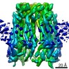
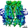
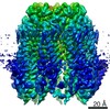
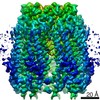
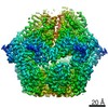
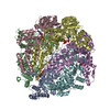
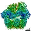
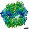
| Title | Cryo-EM structure of wild type human Pannexin 1 channel extracted using SMA 30010 | ||||||||||||
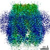
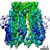
 Map data Map data | Unsharpened map of SMA-wt-hsPANX1 | ||||||||||||
 Sample Sample |
| ||||||||||||
| Function / homology |  Function and homology information Function and homology informationElectric Transmission Across Gap Junctions /  leak channel activity / positive regulation of interleukin-1 alpha production / bleb / wide pore channel activity / gap junction channel activity / gap junction / positive regulation of macrophage cytokine production / oogenesis / response to ATP ...Electric Transmission Across Gap Junctions / leak channel activity / positive regulation of interleukin-1 alpha production / bleb / wide pore channel activity / gap junction channel activity / gap junction / positive regulation of macrophage cytokine production / oogenesis / response to ATP / monoatomic cation transport / The NLRP3 inflammasome / positive regulation of interleukin-1 beta production / response to ischemia / calcium channel activity / calcium ion transport / actin filament binding / cell-cell signaling / scaffold protein binding / protease binding / transmembrane transporter binding / signaling receptor binding / endoplasmic reticulum membrane / structural molecule activity / endoplasmic reticulum / protein-containing complex / membrane / identical protein binding / plasma membrane leak channel activity / positive regulation of interleukin-1 alpha production / bleb / wide pore channel activity / gap junction channel activity / gap junction / positive regulation of macrophage cytokine production / oogenesis / response to ATP ...Electric Transmission Across Gap Junctions / leak channel activity / positive regulation of interleukin-1 alpha production / bleb / wide pore channel activity / gap junction channel activity / gap junction / positive regulation of macrophage cytokine production / oogenesis / response to ATP / monoatomic cation transport / The NLRP3 inflammasome / positive regulation of interleukin-1 beta production / response to ischemia / calcium channel activity / calcium ion transport / actin filament binding / cell-cell signaling / scaffold protein binding / protease binding / transmembrane transporter binding / signaling receptor binding / endoplasmic reticulum membrane / structural molecule activity / endoplasmic reticulum / protein-containing complex / membrane / identical protein binding / plasma membraneSimilarity search - Function | ||||||||||||
| Biological species |  Homo sapiens (human) Homo sapiens (human) | ||||||||||||
| Method | single particle reconstruction / cryo EM / Resolution: 6.04 Å | ||||||||||||
 Authors Authors | Lu W / Du J | ||||||||||||
| Funding support |  United States, 3 items United States, 3 items
| ||||||||||||
 Citation Citation | Journal: Nature / Year: 2020 Title: Structures of human pannexin 1 reveal ion pathways and mechanism of gating. Authors: Zheng Ruan / Ian J Orozco / Juan Du / Wei Lü / Abstract: Pannexin 1 (PANX1) is an ATP-permeable channel with critical roles in a variety of physiological functions such as blood pressure regulation, apoptotic cell clearance and human oocyte development. ...Pannexin 1 (PANX1) is an ATP-permeable channel with critical roles in a variety of physiological functions such as blood pressure regulation, apoptotic cell clearance and human oocyte development. Here we present several structures of human PANX1 in a heptameric assembly at resolutions of up to 2.8 angström, including an apo state, a caspase-7-cleaved state and a carbenoxolone-bound state. We reveal a gating mechanism that involves two ion-conducting pathways. Under normal cellular conditions, the intracellular entry of the wide main pore is physically plugged by the C-terminal tail. Small anions are conducted through narrow tunnels in the intracellular domain. These tunnels connect to the main pore and are gated by a long linker between the N-terminal helix and the first transmembrane helix. During apoptosis, the C-terminal tail is cleaved by caspase, allowing the release of ATP through the main pore. We identified a carbenoxolone-binding site embraced by W74 in the extracellular entrance and a role for carbenoxolone as a channel blocker. We identified a gap-junction-like structure using a glycosylation-deficient mutant, N255A. Our studies provide a solid foundation for understanding the molecular mechanisms underlying the channel gating and inhibition of PANX1 and related large-pore channels. | ||||||||||||
| History |
|
- Structure visualization
| Movie |
Movie viewer |
|---|---|
| Structure viewer | EM map: SurfViewMolmilJmol/JSmol |
| Supplemental images |

- Downloads & links
-EMDB archive
| Map data | emd_21598.map.gz | 92.1 MB | EMDB map data format | |
|---|---|---|---|---|
| Header (meta data) | emd-21598-v30.xmlemd-21598.xml | 13.9 KB 13.9 KB | Display Display | EMDB header |
| Images |  emd_21598.png emd_21598.png | 128.5 KB | ||
| Archive directory |  http://ftp.pdbj.org/pub/emdb/structures/EMD-21598ftp://ftp.pdbj.org/pub/emdb/structures/EMD-21598 http://ftp.pdbj.org/pub/emdb/structures/EMD-21598ftp://ftp.pdbj.org/pub/emdb/structures/EMD-21598 | HTTPS FTP |
-Related structure data
| Related structure data |  6wbfC 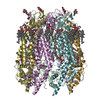 6wbgC 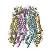 6wbiC 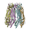 6wbkC 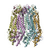 6wblC 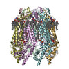 6wbmC 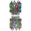 6wbnC C: citing same article ( |
|---|---|
| Similar structure data |


-Links
| EMDB pages | EMDB (EBI/PDBe) / EMDataResource |
|---|---|
| Related items in Molecule of the Month |


-Map
| File | Download / File: emd_21598.map.gz / Format: CCP4 / Size: 103 MB / Type: IMAGE STORED AS FLOATING POINT NUMBER (4 BYTES) | ||||||||||||||||||||||||||||||||||||||||||||||||||||||||||||
|---|---|---|---|---|---|---|---|---|---|---|---|---|---|---|---|---|---|---|---|---|---|---|---|---|---|---|---|---|---|---|---|---|---|---|---|---|---|---|---|---|---|---|---|---|---|---|---|---|---|---|---|---|---|---|---|---|---|---|---|---|---|
| Annotation | Unsharpened map of SMA-wt-hsPANX1 | ||||||||||||||||||||||||||||||||||||||||||||||||||||||||||||
| Voxel size | X=Y=Z: 0.812 Å | ||||||||||||||||||||||||||||||||||||||||||||||||||||||||||||
| Density |
| ||||||||||||||||||||||||||||||||||||||||||||||||||||||||||||
| Symmetry | Space group: 1 | ||||||||||||||||||||||||||||||||||||||||||||||||||||||||||||
| Details | EMDB XML:
CCP4 map header:
| ||||||||||||||||||||||||||||||||||||||||||||||||||||||||||||
-Supplemental data
- Sample components
-Entire : Wild type human Pannexin 1 channel
| Entire | Name: Wild type human Pannexin 1 channel |
|---|---|
| Components |
|
-Supramolecule #1: Wild type human Pannexin 1 channel
| Supramolecule | Name: Wild type human Pannexin 1 channel / type: complex / ID: 1 / Parent: 0 / Macromolecule list: all |
|---|---|
| Source (natural) | Organism: Homo sapiens (human) |
| Recombinant expression | Organism: Homo sapiens (human) |
-Macromolecule #1: Pannexin-1
| Macromolecule | Name: Pannexin-1 / type: protein_or_peptide / ID: 1 / Enantiomer: LEVO |
|---|---|
| Source (natural) | Organism: Homo sapiens (human) |
| Sequence | String: MAIAQLATEY VFSDFLLKEP TEPKFKGLRL ELAVDKMVTC IAVGLPLLLI SLAFAQEISI GTQISCFSP SSFSWRQAAF VDSYCWAAVQ QKNSLQSESG NLPLWLHKFF PYILLLFAIL L YLPPLFWR FAAAPHICSD LKFIMEELDK VYNRAIKAAK SARDLDMRDG ...String: MAIAQLATEY VFSDFLLKEP TEPKFKGLRL ELAVDKMVTC IAVGLPLLLI SLAFAQEISI GTQISCFSP SSFSWRQAAF VDSYCWAAVQ QKNSLQSESG NLPLWLHKFF PYILLLFAIL L YLPPLFWR FAAAPHICSD LKFIMEELDK VYNRAIKAAK SARDLDMRDG ACSVPGVTEN LG QSLWEVS ESHFKYPIVE QYLKTKKNSN NLIIKYISCR LLTLIIILLA CIYLGYYFSL SSL SDEFVC SIKSGILRND STVPDQFQCK LIAVGIFQLL SVINLVVYVL LAPVVVYTLF VPFR QKTDV LKVYEILPTF DVLHFKSEGY NDLSLYNLFL EENISEVKSY KCLKVLENIK SSGQG IDPM LLLTNLGMIK MDVVDGKTPM SAEMREEQGN QTAELQGMNI DSETKANNGE KNARQR LLD SSC |
-Experimental details
-Structure determination
| Method | cryo EM |
|---|---|
 Processing Processing | single particle reconstruction |
| Aggregation state | particle |
-Sample preparation
| Concentration | 7 mg/mL |
|---|---|
| Buffer | pH: 8 |
| Grid | Details: unspecified |
| Vitrification | Cryogen name: ETHANE / Chamber humidity: 100 % / Chamber temperature: 291.15 K / Instrument: FEI VITROBOT MARK III |
- Electron microscopy
| Microscope | FEI TITAN KRIOS |
|---|---|
| Electron beam | Acceleration voltage: 300 kV / Electron source: FIELD EMISSION GUN |
| Electron optics | Illumination mode: FLOOD BEAM / Imaging mode: BRIGHT FIELDBright-field microscopy |
| Image recording | Film or detector model: GATAN K2 SUMMIT (4k x 4k) / Detector mode: SUPER-RESOLUTION / Average exposure time: 8.0 sec. / Average electron dose: 49.6 e/Å2 |
| Experimental equipment |  Model: Titan Krios / Image courtesy: FEI Company |
-Image processing
| Particle selection | Number selected: 1098021 |
|---|---|
| CTF correction | Software - Name: Gctf (ver. 1.06) |
| Startup model | Type of model: OTHER / Details: cryoSPARC ab-initio reconstruction |
| Initial angle assignment | Type: MAXIMUM LIKELIHOOD / Software - Name: RELION (ver. 3.1b) |
| Final 3D classification | Number classes: 10 / Software - Name: RELION (ver. 3.1b) |
| Final angle assignment | Type: MAXIMUM LIKELIHOOD / Software - Name: RELION (ver. 3.1b) |
| Final reconstruction | Applied symmetry - Point group: C7 (7 fold cyclic) / Resolution.type: BY AUTHOR / Resolution: 6.04 Å / Resolution method: FSC 0.143 CUT-OFF / Software - Name: RELION (ver. 3.1b) / Number images used: 125025 |
-Atomic model buiding 1
| Refinement | Space: REAL / Protocol: AB INITIO MODEL |
|---|
