 Movie
Movie Controller

[English] 日本語
 Yorodumi
- EMDB-2153: Electron tomography of Saccharomyces cerevisiae kinetochore fragm... -
+ Open data

- Basic information
| Entry | Database: EMDB / ID: EMD-2153 | |||||||||
|---|---|---|---|---|---|---|---|---|---|---|
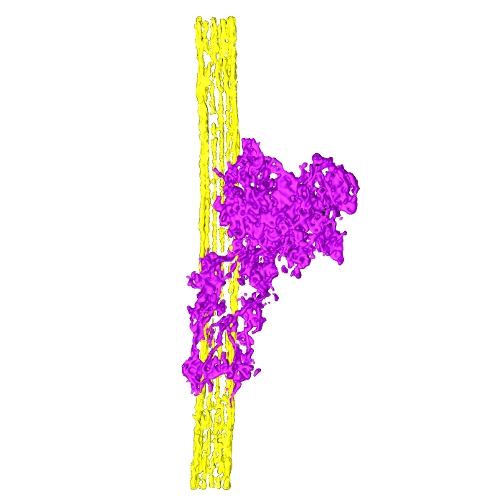
| Title | Electron tomography of Saccharomyces cerevisiae kinetochore fragment on a taxol stabilized microtubule | |||||||||
 Map data Map data | Tomogram of S cerevisiae kinetochore fragment on a taxol-stabilized microtubule | |||||||||
 Sample Sample |
| |||||||||
 Keywords Keywords |  Kinetochore / microtubule / dam1 / ndc80 / KMN / taxol / Saccharomyces cerevisiae Kinetochore / microtubule / dam1 / ndc80 / KMN / taxol / Saccharomyces cerevisiae | |||||||||
| Biological species |  Saccharomyces cerevisiae (brewer's yeast) Saccharomyces cerevisiae (brewer's yeast) | |||||||||
| Method | electron tomography / negative staining | |||||||||
 Authors Authors | Gonen S / Akiyoshi B / Iadanza MG / Shi D / Duggan N / Biggins S / Gonen T | |||||||||
 Citation Citation | Journal: Nat Struct Mol Biol / Year: 2012 Title: The structure of purified kinetochores reveals multiple microtubule-attachment sites. Authors: Shane Gonen / Bungo Akiyoshi / Matthew G Iadanza / Dan Shi / Nicole Duggan / Sue Biggins / Tamir Gonen /  Abstract: Chromosomes must be accurately partitioned to daughter cells to prevent aneuploidy, a hallmark of many tumors and birth defects. Kinetochores are the macromolecular machines that segregate ...Chromosomes must be accurately partitioned to daughter cells to prevent aneuploidy, a hallmark of many tumors and birth defects. Kinetochores are the macromolecular machines that segregate chromosomes by maintaining load-bearing attachments to the dynamic tips of microtubules. Here, we present the structure of isolated budding-yeast kinetochore particles, as visualized by EM and electron tomography of negatively stained preparations. The kinetochore appears as an ~126-nm particle containing a large central hub surrounded by multiple outer globular domains. In the presence of microtubules, some particles also have a ring that encircles the microtubule. Our data, showing that kinetochores bind to microtubules via multivalent attachments, lay the foundation to uncover the key mechanical and regulatory mechanisms by which kinetochores control chromosome segregation and cell division. | |||||||||
| History |
|
- Structure visualization
| Movie |
 Movie viewer Movie viewer |
|---|---|
| Structure viewer | EM map: SurfViewMolmilJmol/JSmol |
| Supplemental images |
- Downloads & links
-EMDB archive
| Map data | emd_2153.map.gz | 67.4 MB | EMDB map data format | |
|---|---|---|---|---|
| Header (meta data) | emd-2153-v30.xmlemd-2153.xml | 9.4 KB 9.4 KB | Display Display | EMDB header |
| Images |  emd_2153.jpg emd_2153.jpg | 56.5 KB | ||
| Archive directory |  http://ftp.pdbj.org/pub/emdb/structures/EMD-2153ftp://ftp.pdbj.org/pub/emdb/structures/EMD-2153 http://ftp.pdbj.org/pub/emdb/structures/EMD-2153ftp://ftp.pdbj.org/pub/emdb/structures/EMD-2153 | HTTPS FTP |
-Related structure data
-Links
| EMDB pages | EMDB (EBI/PDBe) / EMDataResource |
|---|---|
| Related items in Molecule of the Month |

-Map
| File | Download / File: emd_2153.map.gz / Format: CCP4 / Size: 97.8 MB / Type: IMAGE STORED AS SIGNED BYTE | ||||||||||||||||||||||||||||||||||||||||||||||||||||||||||||||||||||
|---|---|---|---|---|---|---|---|---|---|---|---|---|---|---|---|---|---|---|---|---|---|---|---|---|---|---|---|---|---|---|---|---|---|---|---|---|---|---|---|---|---|---|---|---|---|---|---|---|---|---|---|---|---|---|---|---|---|---|---|---|---|---|---|---|---|---|---|---|---|
| Annotation | Tomogram of S cerevisiae kinetochore fragment on a taxol-stabilized microtubule | ||||||||||||||||||||||||||||||||||||||||||||||||||||||||||||||||||||
| Voxel size | X=Y=Z: 4.3 Å | ||||||||||||||||||||||||||||||||||||||||||||||||||||||||||||||||||||
| Density |
| ||||||||||||||||||||||||||||||||||||||||||||||||||||||||||||||||||||
| Symmetry | Space group: 1 | ||||||||||||||||||||||||||||||||||||||||||||||||||||||||||||||||||||
| Details | EMDB XML:
CCP4 map header:
| ||||||||||||||||||||||||||||||||||||||||||||||||||||||||||||||||||||
-Supplemental data
- Sample components
-Entire : Saccharomyces cerevisiae kinetochore fragment on a taxol stabiliz...
| Entire | Name: Saccharomyces cerevisiae kinetochore fragment on a taxol stabilized microtubule |
|---|---|
| Components |
|
-Supramolecule #1000: Saccharomyces cerevisiae kinetochore fragment on a taxol stabiliz...
| Supramolecule | Name: Saccharomyces cerevisiae kinetochore fragment on a taxol stabilized microtubule type: sample / ID: 1000 Details: fragment of native kinetochore, individual components not identified Number unique components: 2 |
|---|
-Supramolecule #1: Microtubule
| Supramolecule | Name: Microtubule / type: organelle_or_cellular_component / ID: 1 / Details: stabilized with taxol / Number of copies: 1 / Recombinant expression: No / Database: NCBI |
|---|---|
| Source (natural) | Organism: Saccharomyces cerevisiae (brewer's yeast) / Strain: SBY8253 / synonym: Baker's Yeast / Location in cell: Cytoplasm |
-Supramolecule #2: Kinetochore fragment
| Supramolecule | Name: Kinetochore fragment / type: organelle_or_cellular_component / ID: 2 / Details: individual components not identified / Number of copies: 1 / Recombinant expression: No / Database: NCBI |
|---|---|
| Source (natural) | Organism: Saccharomyces cerevisiae (brewer's yeast) / Strain: SBY8253 / synonym: Baker's Yeast / Location in cell: Cytoplasm |
-Experimental details
-Structure determination
| Method | negative staining |
|---|---|
 Processing Processing | electron tomography |
-Sample preparation
| Buffer | pH: 8 Details: 25 mM HEPES, 0.2 mM MgCl2, 0.1 mM EDTA, 0.5 mM EGTA, 150 mM KCl, 15% (v/v) Glycerol, 0.1% (v/v) NP-40 |
|---|---|
| Staining | Type: NEGATIVE Details: Sample applied to grid for 20 sec, washed with one drop of ultrapure water and two drops of 0.75% uranyl formate. Anti-mouse IgG-gold fiducial markers added and second layer of carbon applied by evaporation. |
| Grid | Details: Gilder 400 mesh copper grid, carbon coated by evaporation. Positive discharge. |
| Vitrification | Cryogen name: NONE / Instrument: OTHER |
- Electron microscopy
| Microscope | FEI TECNAI 12 |
|---|---|
| Electron beam | Acceleration voltage: 120 kV / Electron source: LAB6 |
| Electron optics | Calibrated magnification: 52000 / Illumination mode: FLOOD BEAM / Imaging mode: BRIGHT FIELDBright-field microscopy / Nominal defocus max: 2.0 µm / Nominal defocus min: 2.0 µm |
| Sample stage | Specimen holder model: OTHER / Tilt series - Axis1 - Min angle: -70 ° / Tilt series - Axis1 - Max angle: 70 ° / Tilt series - Axis1 - Angle increment: 2 ° |
| Date | Jan 6, 2012 |
| Image recording | Category: CCD / Film or detector model: GENERIC GATAN (4k x 4k) / Number real images: 100 |
-Image processing
| Final reconstruction | Software - Name: Amira, IMOD / Number images used: 100 |
|---|
