 Movie
Movie Controller

[English] 日本語
 Yorodumi
- EMDB-2052: Capsid structure and its Stability at the Late Stages of Bacterio... -
+ Open data

- Basic information
| Entry | Database: EMDB / ID: EMD-2052 | |||||||||
|---|---|---|---|---|---|---|---|---|---|---|
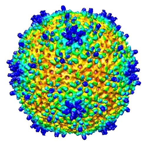
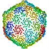
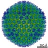
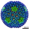
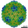
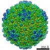
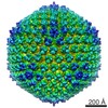
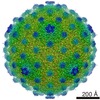
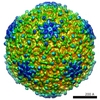
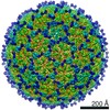
| Title | Capsid structure and its Stability at the Late Stages of Bacteriophage SPP1 Assembly | |||||||||
 Map data Map data | Reconstruction of an SPP1 capsid after DNA ejection | |||||||||
 Sample Sample |
| |||||||||
 Keywords Keywords | Bacteriophage Capsid SPP1 | |||||||||
| Biological species |  Bacillus phage SPP1 (virus) Bacillus phage SPP1 (virus) | |||||||||
| Method | single particle reconstruction / cryo EM / Resolution: 10.5 Å | |||||||||
 Authors Authors | White HE / Sherman MB / Brasiles S / Jacquet E / Seavers P / Tavares P / Orlova EV | |||||||||
 Citation Citation | Journal: J Virol / Year: 2012 Title: Capsid structure and its stability at the late stages of bacteriophage SPP1 assembly. Authors: Helen E White / Michael B Sherman / Sandrine Brasilès / Eric Jacquet / Philippa Seavers / Paulo Tavares / Elena V Orlova /  Abstract: The structure of the bacteriophage SPP1 capsid was determined at subnanometer resolution by cryo-electron microscopy and single-particle analysis. The icosahedral capsid is composed of the major ...The structure of the bacteriophage SPP1 capsid was determined at subnanometer resolution by cryo-electron microscopy and single-particle analysis. The icosahedral capsid is composed of the major capsid protein gp13 and the auxiliary protein gp12, which are organized in a T=7 lattice. DNA is arranged in layers with a distance of ~24.5 Å. gp12 forms spikes that are anchored at the center of gp13 hexamers. In a gp12-deficient mutant, the centers of hexamers are closed by loops of gp13 coming together to protect the SPP1 genome from the outside environment. The HK97-like fold was used to build a pseudoatomic model of gp13. Its structural organization remains unchanged upon tail binding and following DNA release. gp13 exhibits enhanced thermostability in the DNA-filled capsid. A remarkable convergence between the thermostability of the capsid and those of the other virion components was found, revealing that the overall architecture of the SPP1 infectious particle coevolved toward high robustness. | |||||||||
| History |
|
- Structure visualization
| Movie |
 Movie viewer Movie viewer |
|---|---|
| Structure viewer | EM map: SurfViewMolmilJmol/JSmol |
| Supplemental images |
- Downloads & links
-EMDB archive
| Map data | emd_2052.map.gz | 119 MB | EMDB map data format | |
|---|---|---|---|---|
| Header (meta data) | emd-2052-v30.xmlemd-2052.xml | 7.8 KB 7.8 KB | Display Display | EMDB header |
| Images |  EMD-2052.jpg EMD-2052.jpg | 226.5 KB | ||
| Archive directory |  http://ftp.pdbj.org/pub/emdb/structures/EMD-2052ftp://ftp.pdbj.org/pub/emdb/structures/EMD-2052 http://ftp.pdbj.org/pub/emdb/structures/EMD-2052ftp://ftp.pdbj.org/pub/emdb/structures/EMD-2052 | HTTPS FTP |
-Validation report
| Summary document | emd_2052_validation.pdf.gz | 291.4 KB | Display | EMDB validaton report |
|---|---|---|---|---|
| Full document | emd_2052_full_validation.pdf.gz | 290.5 KB | Display | |
| Data in XML | emd_2052_validation.xml.gz | 8.3 KB | Display | |
| Arichive directory | https://ftp.pdbj.org/pub/emdb/validation_reports/EMD-2052ftp://ftp.pdbj.org/pub/emdb/validation_reports/EMD-2052 | HTTPS FTP |
-Related structure data
-Links
| EMDB pages | EMDB (EBI/PDBe) / EMDataResource |
|---|
-Map
| File | Download / File: emd_2052.map.gz / Format: CCP4 / Size: 500 MB / Type: IMAGE STORED AS FLOATING POINT NUMBER (4 BYTES) | ||||||||||||||||||||||||||||||||||||||||||||||||||||||||||||||||||||
|---|---|---|---|---|---|---|---|---|---|---|---|---|---|---|---|---|---|---|---|---|---|---|---|---|---|---|---|---|---|---|---|---|---|---|---|---|---|---|---|---|---|---|---|---|---|---|---|---|---|---|---|---|---|---|---|---|---|---|---|---|---|---|---|---|---|---|---|---|---|
| Annotation | Reconstruction of an SPP1 capsid after DNA ejection | ||||||||||||||||||||||||||||||||||||||||||||||||||||||||||||||||||||
| Voxel size | X: 1.491 Å / Y: 1.49 Å / Z: 1.49 Å | ||||||||||||||||||||||||||||||||||||||||||||||||||||||||||||||||||||
| Density |
| ||||||||||||||||||||||||||||||||||||||||||||||||||||||||||||||||||||
| Symmetry | Space group: 1 | ||||||||||||||||||||||||||||||||||||||||||||||||||||||||||||||||||||
| Details | EMDB XML:
CCP4 map header:
| ||||||||||||||||||||||||||||||||||||||||||||||||||||||||||||||||||||
-Supplemental data
- Sample components
-Entire : Capsid of SPP1
| Entire | Name: Capsid of SPP1 |
|---|---|
| Components |
|
-Supramolecule #1000: Capsid of SPP1
| Supramolecule | Name: Capsid of SPP1 / type: sample / ID: 1000 / Oligomeric state: Icosahedron / Number unique components: 1 |
|---|
-Supramolecule #1: Bacillus phage SPP1
| Supramolecule | Name: Bacillus phage SPP1 / type: virus / ID: 1 / NCBI-ID: 10724 / Sci species name: Bacillus phage SPP1 / Virus type: OTHER / Virus isolate: SPECIES / Virus enveloped: No / Virus empty: Yes |
|---|---|
| Host (natural) | Organism:  |
| Virus shell | Shell ID: 1 / Diameter: 600 Å / T number (triangulation number): 7 |
-Experimental details
-Structure determination
| Method | cryo EM |
|---|---|
 Processing Processing | single particle reconstruction |
| Aggregation state | particle |
-Sample preparation
| Grid | Details: holey carbon film grids |
|---|---|
| Vitrification | Cryogen name: ETHANE / Instrument: FEI VITROBOT MARK I |
- Electron microscopy
| Microscope | JEOL 2200FS |
|---|---|
| Details | Low-dose imaging procedure |
| Date | Apr 6, 2005 |
| Image recording | Category: FILM / Film or detector model: KODAK SO-163 FILM / Digitization - Scanner: ZEISS SCAI / Digitization - Sampling interval: 7 µm / Number real images: 94 / Average electron dose: 20 e/Å2 |
| Electron beam | Acceleration voltage: 200 kV / Electron source:  FIELD EMISSION GUN FIELD EMISSION GUN |
| Electron optics | Illumination mode: FLOOD BEAM / Imaging mode: BRIGHT FIELD / Nominal magnification: 50000 |
| Sample stage | Specimen holder model: GATAN LIQUID NITROGEN |
-Image processing
| CTF correction | Details: Phase flipping |
|---|---|
| Final reconstruction | Applied symmetry - Point group: I (icosahedral) / Algorithm: OTHER / Resolution.type: BY AUTHOR / Resolution: 10.5 Å / Resolution method: FSC 0.5 CUT-OFF / Software - Name: Imagic / Number images used: 1200 |
