Movie
Movie Controller

[English] 日本語
 Yorodumi
- EMDB-2010: 3D reconstruction of a translating yeast 80S ribosome in complex ... -
+ Open data

- Basic information
| Entry | Database: EMDB / ID: EMD-2010 | |||||||||
|---|---|---|---|---|---|---|---|---|---|---|
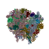
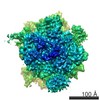
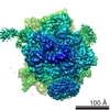
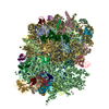
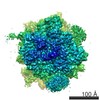
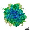
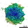
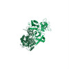
| Title | 3D reconstruction of a translating yeast 80S ribosome in complex with Dom34p and Rli1p | |||||||||
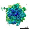
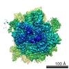
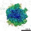
 Map data Map data | This map represents a stalled yeast 80S ribosome in complex with Dom34 and Rli1 before sorting for P-site tRNA. | |||||||||
 Sample Sample |
| |||||||||
 Keywords Keywords |  translation / ribosome recycling / no-go mRNA decay translation / ribosome recycling / no-go mRNA decay | |||||||||
| Function / homology |  Function and homology information Function and homology informationDom34-Hbs1 complex / RNA surveillance / nuclear-transcribed mRNA catabolic process, no-go decay / nuclear-transcribed mRNA catabolic process, non-stop decay / nonfunctional rRNA decay / ribosome disassembly / mTORC1-mediated signalling / Formation of the ternary complex, and subsequently, the 43S complex / Translation initiation complex formation / Ribosomal scanning and start codon recognition ...Dom34-Hbs1 complex / RNA surveillance / nuclear-transcribed mRNA catabolic process, no-go decay / nuclear-transcribed mRNA catabolic process, non-stop decay / nonfunctional rRNA decay / ribosome disassembly / mTORC1-mediated signalling / Formation of the ternary complex, and subsequently, the 43S complex / Translation initiation complex formation / Ribosomal scanning and start codon recognition / ribosomal subunit export from nucleus / Major pathway of rRNA processing in the nucleolus and cytosol / SRP-dependent cotranslational protein targeting to membrane / 90S preribosome / GTP hydrolysis and joining of the 60S ribosomal subunit / Formation of a pool of free 40S subunits / Nonsense Mediated Decay (NMD) independent of the Exon Junction Complex (EJC) / Nonsense Mediated Decay (NMD) enhanced by the Exon Junction Complex (EJC) / ribosomal small subunit binding / positive regulation of translational initiation / L13a-mediated translational silencing of Ceruloplasmin expression / rescue of stalled ribosome / translational termination / maturation of SSU-rRNA from tricistronic rRNA transcript (SSU-rRNA, 5.8S rRNA, LSU-rRNA) / ribosomal large subunit biogenesis / RNA endonuclease activity / translational initiation / translation initiation factor activity / small-subunit processome / meiotic cell cycle / positive regulation of translation / ribosomal large subunit assembly / rRNA processing / cytoplasmic stress granule / cytosolic small ribosomal subunit / large ribosomal subunit rRNA binding / cytoplasmic translation / cytosolic large ribosomal subunit / rRNA binding / structural constituent of ribosome / translation / iron ion binding / cell division / nucleolus / ATP hydrolysis activity / mitochondrion / nucleoplasm / ATP binding / metal ion binding / nucleus / cytosol / cytoplasmSimilarity search - Function | |||||||||
| Biological species |  Saccharomyces cerevisiae (brewer's yeast) Saccharomyces cerevisiae (brewer's yeast) | |||||||||
| Method | single particle reconstruction / cryo EM / Resolution: 7.2 Å | |||||||||
 Authors Authors | Becker T / Franckenberg S / Wickles S / Shoemaker CJ / Anger AM / Armache J-P / Sieber H / Ungewickell C / Berninghausen O / Daberkow I ...Becker T / Franckenberg S / Wickles S / Shoemaker CJ / Anger AM / Armache J-P / Sieber H / Ungewickell C / Berninghausen O / Daberkow I / Karcher A / Thomm M / Hopfner K-P / Green R / Beckmann R | |||||||||
 Citation Citation | Journal: Nature / Year: 2012 Title: Structural basis of highly conserved ribosome recycling in eukaryotes and archaea. Authors: Thomas Becker / Sibylle Franckenberg / Stephan Wickles / Christopher J Shoemaker / Andreas M Anger / Jean-Paul Armache / Heidemarie Sieber / Charlotte Ungewickell / Otto Berninghausen / Ingo ...Authors: Thomas Becker / Sibylle Franckenberg / Stephan Wickles / Christopher J Shoemaker / Andreas M Anger / Jean-Paul Armache / Heidemarie Sieber / Charlotte Ungewickell / Otto Berninghausen / Ingo Daberkow / Annette Karcher / Michael Thomm / Karl-Peter Hopfner / Rachel Green / Roland Beckmann /  Abstract: Ribosome-driven protein biosynthesis is comprised of four phases: initiation, elongation, termination and recycling. In bacteria, ribosome recycling requires ribosome recycling factor and elongation ...Ribosome-driven protein biosynthesis is comprised of four phases: initiation, elongation, termination and recycling. In bacteria, ribosome recycling requires ribosome recycling factor and elongation factor G, and several structures of bacterial recycling complexes have been determined. In the eukaryotic and archaeal kingdoms, however, recycling involves the ABC-type ATPase ABCE1 and little is known about its structural basis. Here we present cryo-electron microscopy reconstructions of eukaryotic and archaeal ribosome recycling complexes containing ABCE1 and the termination factor paralogue Pelota. These structures reveal the overall binding mode of ABCE1 to be similar to canonical translation factors. Moreover, the iron-sulphur cluster domain of ABCE1 interacts with and stabilizes Pelota in a conformation that reaches towards the peptidyl transferase centre, thus explaining how ABCE1 may stimulate peptide-release activity of canonical termination factors. Using the mechanochemical properties of ABCE1, a conserved mechanism in archaea and eukaryotes is suggested that couples translation termination to recycling, and eventually to re-initiation. | |||||||||
| History |
|
- Structure visualization
| Movie |
Movie viewer |
|---|---|
| Structure viewer | EM map: SurfViewMolmilJmol/JSmol |
| Supplemental images |

- Downloads & links
-EMDB archive
| Map data | emd_2010.map.gz | 23.8 MB | EMDB map data format | |
|---|---|---|---|---|
| Header (meta data) | emd-2010-v30.xmlemd-2010.xml | 13 KB 13 KB | Display Display | EMDB header |
| Images |  EMBD_2010.gif EMBD_2010.gif | 238 KB | ||
| Archive directory |  http://ftp.pdbj.org/pub/emdb/structures/EMD-2010ftp://ftp.pdbj.org/pub/emdb/structures/EMD-2010 http://ftp.pdbj.org/pub/emdb/structures/EMD-2010ftp://ftp.pdbj.org/pub/emdb/structures/EMD-2010 | HTTPS FTP |
-Related structure data
| Related structure data | 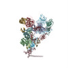 3j16MC 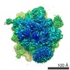 2008C  2009C 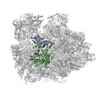 3j15C M: atomic model generated by this map C: citing same article ( |
|---|---|
| Similar structure data |
-Links
| EMDB pages | EMDB (EBI/PDBe) / EMDataResource |
|---|---|
| Related items in Molecule of the Month |


-Map
| File | Download / File: emd_2010.map.gz / Format: CCP4 / Size: 185.7 MB / Type: IMAGE STORED AS FLOATING POINT NUMBER (4 BYTES) | ||||||||||||||||||||||||||||||||||||||||||||||||||||||||||||||||||||
|---|---|---|---|---|---|---|---|---|---|---|---|---|---|---|---|---|---|---|---|---|---|---|---|---|---|---|---|---|---|---|---|---|---|---|---|---|---|---|---|---|---|---|---|---|---|---|---|---|---|---|---|---|---|---|---|---|---|---|---|---|---|---|---|---|---|---|---|---|---|
| Annotation | This map represents a stalled yeast 80S ribosome in complex with Dom34 and Rli1 before sorting for P-site tRNA. | ||||||||||||||||||||||||||||||||||||||||||||||||||||||||||||||||||||
| Voxel size | X=Y=Z: 1.2375 Å | ||||||||||||||||||||||||||||||||||||||||||||||||||||||||||||||||||||
| Density |
| ||||||||||||||||||||||||||||||||||||||||||||||||||||||||||||||||||||
| Symmetry | Space group: 1 | ||||||||||||||||||||||||||||||||||||||||||||||||||||||||||||||||||||
| Details | EMDB XML:
CCP4 map header:
| ||||||||||||||||||||||||||||||||||||||||||||||||||||||||||||||||||||
-Supplemental data
- Sample components
-Entire : Saccharomyces cerevisiae 80S ribosome stalled by a synthetic stem...
| Entire | Name: Saccharomyces cerevisiae 80S ribosome stalled by a synthetic stem-loop mRNA in complex with Dom34p and Rli1p |
|---|---|
| Components |
|
-Supramolecule #1000: Saccharomyces cerevisiae 80S ribosome stalled by a synthetic stem...
| Supramolecule | Name: Saccharomyces cerevisiae 80S ribosome stalled by a synthetic stem-loop mRNA in complex with Dom34p and Rli1p type: sample / ID: 1000 Oligomeric state: One 80S ribosome binds one Dom34p and one Rli1p Number unique components: 3 |
|---|
-Supramolecule #1: S. cerevisiae 80S ribosome
| Supramolecule | Name: S. cerevisiae 80S ribosome / type: complex / ID: 1 / Name.synonym: Baker's yeast 80S ribosome / Recombinant expression: No / Ribosome-details: ribosome-eukaryote: ALL |
|---|---|
| Source (natural) | Organism: Saccharomyces cerevisiae (brewer's yeast) / synonym: Baker's yeast |
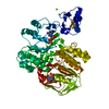
-Macromolecule #1: Rli1p
| Macromolecule | Name: Rli1p / type: protein_or_peptide / ID: 1 / Name.synonym: ABCE1 / Oligomeric state: Monomer / Recombinant expression: Yes |
|---|---|
| Source (natural) | Organism: Saccharomyces cerevisiae (brewer's yeast) / synonym: Baker's yeast |
| Recombinant expression | Organism: Saccharomyces cerevisiae (brewer's yeast) / Recombinant plasmid: pYES |
-Macromolecule #2: Dom34p
| Macromolecule | Name: Dom34p / type: protein_or_peptide / ID: 2 / Name.synonym: Pelota / Oligomeric state: Monomer / Recombinant expression: Yes |
|---|---|
| Source (natural) | Organism: Saccharomyces cerevisiae (brewer's yeast) / synonym: Baker's Yeast |
| Recombinant expression | Organism: Saccharomyces cerevisiae (brewer's yeast) / Recombinant plasmid: pET28 |
-Experimental details
-Structure determination
| Method | cryo EM |
|---|---|
 Processing Processing | single particle reconstruction |
| Aggregation state | particle |
-Sample preparation
| Buffer | pH: 7 Details: 20 mM Tris/HCl pH 7.0, 150 mM KOAc, 10 mM Mg(OAc)2, 1.5 mM DTT, 0.005% Nikkol, 0.01 mg/ml Cycloheximide, 0.3% (w/v) Digitonin, 0.5 mM ADPNP |
|---|---|
| Grid | Details: Quantifoil grids (3/3) with 2 nm carbon on top |
| Vitrification | Cryogen name: ETHANE / Chamber humidity: 95 % / Instrument: OTHER / Details: Vitrification instrument: Vitrobot Method: Blot for 10 seconds before plunging, use 2 layer of filter paper |
- Electron microscopy
| Microscope | FEI TITAN KRIOS |
|---|---|
| Electron beam | Acceleration voltage: 300 kV / Electron source: FIELD EMISSION GUN |
| Electron optics | Illumination mode: FLOOD BEAM / Imaging mode: BRIGHT FIELDBright-field microscopy / Cs: 2.7 mm / Nominal defocus max: 4.5 µm / Nominal defocus min: 1.4 µm / Nominal magnification: 75000 |
| Sample stage | Specimen holder: Dual tilt autoloader cartridge / Specimen holder model: OTHER |
| Details | Final magnification of the object on the CCD image is 128200 |
| Image recording | Category: CCD / Film or detector model: FEI EAGLE (4k x 4k) / Digitization - Sampling interval: 15 µm / Number real images: 5610 / Average electron dose: 25 e/Å2 / Bits/pixel: 16 |
| Experimental equipment |  Model: Titan Krios / Image courtesy: FEI Company |
-Image processing
| CTF correction | Details: Wiener Filter |
|---|---|
| Final angle assignment | Details: SPIDER |
| Final reconstruction | Applied symmetry - Point group: C1 (asymmetric) / Algorithm: OTHER / Resolution.type: BY AUTHOR / Resolution: 7.2 Å / Resolution method: FSC 0.5 CUT-OFF / Software - Name: SPIDER Details: sorting for ribosome conformation and ligand presence was performed. This map represents the step after sorting for ligand presence but before sorting for p_site tRNA. Number images used: 45700 |
| Details | mammalian Sec61 complex was added to saturate hydrophobic nascent polypeptide chains |
-Atomic model buiding 1
| Initial model | PDB ID: Chain - Chain ID: 0 |
|---|---|
| Software | Name: MDFF |
| Details | PDBEntryID_givenInChain. Protocol: rigid body followed by molecular dynamics flexible fitting. rigid body fitting of individual domains followed by molecular dynamics flexible fitting |
| Refinement | Space: REAL / Protocol: FLEXIBLE FIT |
| Output model | PDB-3j16: |
-Atomic model buiding 2
| Initial model | PDB ID: Chain - Chain ID: A |
|---|---|
| Software | Name: MDFF |
| Details | PDBEntryID_givenInChain. Protocol: rigid body followed by molecular dynamics flexible fitting. rigid body followed by molecular dynamics flexible fitting |
| Refinement | Space: REAL / Protocol: FLEXIBLE FIT |
| Output model | PDB-3j16: |