 Movie
Movie Controller

[English] 日本語
 Yorodumi
- PDB-1yxn: Pseudo-atomic model of a fiberless isometric variant of bacteriop... -
+ Open data

- Basic information
| Entry | Database: PDB / ID: 1yxn | ||||||
|---|---|---|---|---|---|---|---|
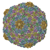
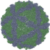
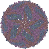
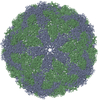
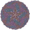
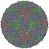
| Title | Pseudo-atomic model of a fiberless isometric variant of bacteriophage phi29 | ||||||
 Components Components | Major head protein | ||||||
 Keywords Keywords |  VIRUS / phi29 / capsid / icosahedral virus capsid / hk97 fold / phage / bacterial immuno-globulin / BIG2 / Icosahedral virus VIRUS / phi29 / capsid / icosahedral virus capsid / hk97 fold / phage / bacterial immuno-globulin / BIG2 / Icosahedral virus | ||||||
| Biological species |   Bacillus phage phi29 (virus) Bacillus phage phi29 (virus) | ||||||
| Method | ELECTRON MICROSCOPY / single particle reconstruction / cryo EM / Resolution: 7.9 Å | ||||||
 Authors Authors | Morais, M.C. / Choi, K.H. / Koti, J.S. / Chipman, P.R. / Anderson, D.L. / Rossmann, M.G. | ||||||
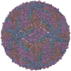
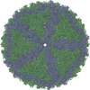
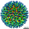
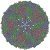
 Citation Citation | Journal: Mol Cell / Year: 2005 Title: Conservation of the capsid structure in tailed dsDNA bacteriophages: the pseudoatomic structure of phi29. Authors: Marc C Morais / Kyung H Choi / Jaya S Koti / Paul R Chipman / Dwight L Anderson / Michael G Rossmann /  Abstract: Bacteriophage phi29 is one of the smallest and simplest known dsDNA phages, making it amenable to structural investigations. The three-dimensional structure of a fiberless, isometric variant has been ...Bacteriophage phi29 is one of the smallest and simplest known dsDNA phages, making it amenable to structural investigations. The three-dimensional structure of a fiberless, isometric variant has been determined to 7.9 A resolution by cryo-electron microscopy (cryo-EM), allowing the identification of alpha helices and beta sheets. Their arrangement indicates that the folds of the phi29 and bacteriophage HK97 capsid proteins are similar except for an additional immunoglobulin-like domain of the phi29 protein. An atomic model that incorporates these two domains fits well into the cryo-EM density of the T = 3, fiberless isometric phi29 particle, and cryo-EM structures of fibered isometric and fiberless prolate prohead phi29 particles at resolutions of 8.7 A and 12.7 A, respectively. Thus, phi29 joins the growing number of phages that utilize the HK97 capsid structure, suggesting that this protein fold may be as prevalent in capsids of dsDNA phages as the jelly roll fold is in eukaryotic viruses. | ||||||
| History |
| ||||||
| Remark 999 | SEQUENCE The authors state that an alignment between the coordinate C-alpha atoms and the sequence ...SEQUENCE The authors state that an alignment between the coordinate C-alpha atoms and the sequence is not possible. The following is the one-letter sequence for the protein modeled in this structure, SwissProt entry P13849: MRITFNDVKTSLGITESYDIVNAIRNSQGDNFKSYVPLATANNVAEVGAGILINQTVQND FITSLVDRIGLVVIRQVSLNNPLKKFKKGQIPLGRTIEEIYTDITKEKQYDAEEAEQKVF EREMPNVKTLFHERNRQGFYHQTIQDDSLKTAFVSWGNFESFVSSIINAIYNSAEVDEYE YMKLLVDNYYSKGLFTTVKIDEPTSSTGALTEFVKKMRATARKLTLPQGSRDWNSMAVRT RSYMEDLHLIIDADLEAELDVDVLAKAFNMNRTDFLGNVTVIDGFASTGLEAVLVDKDWF MVYDNLHKMETVRNPRGLYWNYYYHVWQTLSVSRFANAVAFVSGDVPAVTQVIVSPNIAA VKQGGQQQFTAYVRATNAKDHKVVWSVEGGSTGTAITGDGLLSVSGNEDNQLTVKATVDI GTEDKPKLVVGEAVVSIRPNNASGGAQA |
- Structure visualization
| Movie |
 Movie viewer Movie viewer |
|---|---|
| Structure viewer | Molecule: MolmilJmol/JSmol |
- Downloads & links
-Download
| PDBx/mmCIF format | 1yxn.cif.gz | 37.8 KB | Display | PDBx/mmCIF format |
|---|---|---|---|---|
| PDB format | pdb1yxn.ent.gz | 23.8 KB | Display | PDB format |
| PDBx/mmJSON format | 1yxn.json.gz | Tree view | PDBx/mmJSON format | |
| Others |  Other downloads Other downloads |
-Validation report
| Arichive directory | https://data.pdbj.org/pub/pdb/validation_reports/yx/1yxnftp://data.pdbj.org/pub/pdb/validation_reports/yx/1yxn | HTTPS FTP |
|---|
-Related structure data
| Related structure data | 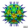 1116MC  1120MC 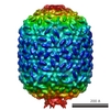 1117C M: map data used to model this data C: citing same article ( |
|---|---|
| Similar structure data |


-Links
 PDBj
PDBj- Assembly
| Deposited unit | 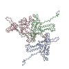
|
|---|---|
| 1 | x 60
|
| 2 |
|
| 3 | x 5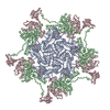
|
| 4 | x 6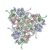
|
| 5 | 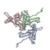
|
| Symmetry | Point symmetry: (Hermann–Mauguin notation: 532 / Schoenflies symbol: I (icosahedral)) |
-Components
| #1: Protein | Mass: 30655.668 Da / Num. of mol.: 3 Source method: isolated from a genetically manipulated source Source: (gene. exp.) Bacillus phage phi29 (virus) / Genus: Phi29-like viruses / Cell line: phi29 / Gene: 8 / Production host:  Bacillus subtilis (bacteria) Bacillus subtilis (bacteria) |
|---|
-Experimental details
-Experiment
| Experiment | Method: ELECTRON MICROSCOPY |
|---|---|
| EM experiment | Aggregation state: PARTICLE / 3D reconstruction method: single particle reconstruction |
- Sample preparation
| Component | Name: Fiberless isometric phi29 capsid / Type: VIRUS Details: The capsid protein assembles a T=3 icosahedral virus shell. The virus particle is a protein shell with icosahedral symmetry. To generate the complete icosahedral particle, apply the ...Details: The capsid protein assembles a T=3 icosahedral virus shell. The virus particle is a protein shell with icosahedral symmetry. To generate the complete icosahedral particle, apply the following 59 matrices to the three chains in the icosahedral asymmetric unit, and combine with the original coordinates of the icosahedral asymmetric unit: Source: NATURAL |
|---|---|
| Source (natural) | Organism: Enterobacteria phage T4 (virus) |
| Details of virus | Host category: BACTERIA / Type: VIRION |
| Natural host | Organism: Bacillus subtilus |
| Buffer solution | Name: tris-HCLTris / pH: 7.8 / Details: tris-HCL |
| Specimen | Conc.: 1 mg/ml / Embedding applied: NO / Shadowing applied: NO / Staining applied: NO / Vitrification applied: YES |
| Specimen support | Details: electron microscopy grid - in vitreous ice |
| Vitrification | Instrument: HOMEMADE PLUNGER / Cryogen name: ETHANE / Details: frozen in liquid ethane |
- Electron microscopy imaging
| Microscopy | Model: FEI/PHILIPS CM200FEG/ST |
|---|---|
| Electron gun | Electron source: FIELD EMISSION GUN / Accelerating voltage: 200 kV / Illumination mode: FLOOD BEAM |
| Electron lens | Mode: BRIGHT FIELDBright-field microscopy / Nominal magnification: 38000 X / Nominal defocus max: 3000 nm / Nominal defocus min: 700 nm / Cs: 2 mm |
| Specimen holder | Temperature: 100 K / Tilt angle max: 0 ° / Tilt angle min: 0 ° |
| Image recording | Electron dose: 20 e/Å2 / Film or detector model: KODAK SO-163 FILM |
- Processing
| EM software |
| ||||||||||||||||||||||||||||
|---|---|---|---|---|---|---|---|---|---|---|---|---|---|---|---|---|---|---|---|---|---|---|---|---|---|---|---|---|---|
| CTF correction | Details: Inverse of CTF was applied to images. Both phases and amplitudes were corrected. | ||||||||||||||||||||||||||||
| Symmetry | Point symmetry: I (icosahedral) | ||||||||||||||||||||||||||||
| 3D reconstruction | Method: 3D reconstructions were calculated using the Fourier-Bessel method. Initial orientations were found via common-lines, improved using model based polar Fourier transform methods, and finally ...Method: 3D reconstructions were calculated using the Fourier-Bessel method. Initial orientations were found via common-lines, improved using model based polar Fourier transform methods, and finally refined by minimizing the vector difference between between structure factors calculated from a particle image and those from a central section of the Fourier transform of the model. Resolution: 7.9 Å / Num. of particles: 5922 / Nominal pixel size: 1.8421 Å Details: Software used included P3DR (3D reconstructions), PFT (initial orientation improvement), and POR (final orientation refinement. Symmetry type: POINT | ||||||||||||||||||||||||||||
| Atomic model building | Protocol: RIGID BODY FIT / Space: REAL Target criteria: rigid body refinement in real space against laplacian filtered EM density, using the program COLORES in the package SITUS. Each molecule in the T=3 asymmetric unit was refined separately. Details: METHOD--6D search for each symmetry related molecule in the icosahedral asymmetric unit REFINEMENT PROTOCOL--rigid body | ||||||||||||||||||||||||||||
| Refinement step | Cycle: LAST
|
