positive regulation of axon guidance / microtubule-based process / Hydrolases; Acting on acid anhydrides; Acting on GTP to facilitate cellular and subcellular movement / structural constituent of cytoskeleton / microtubule cytoskeleton organization / microtubule cytoskeleton / mitotic cell cycle / nervous system development / microtubule / hydrolase activity ...positive regulation of axon guidance / microtubule-based process / Hydrolases; Acting on acid anhydrides; Acting on GTP to facilitate cellular and subcellular movement / structural constituent of cytoskeleton / microtubule cytoskeleton organization / microtubule cytoskeleton / mitotic cell cycle / nervous system development / microtubule / hydrolase activity / protein heterodimerization activity / GTPase activity / GTP binding / metal ion binding / cytoplasm Similarity search - Function
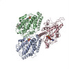
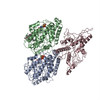
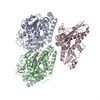
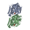
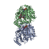
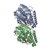
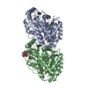
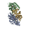
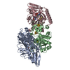
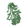
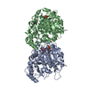
Journal: Science / Year: 2004 Title: The binding mode of epothilone A on alpha,beta-tubulin by electron crystallography. Authors: James H Nettles / Huilin Li / Ben Cornett / Joseph M Krahn / James P Snyder / Kenneth H Downing / Abstract: The structure of epothilone A, bound to alpha,beta-tubulin in zinc-stabilized sheets, was determined by a combination of electron crystallography at 2.89 angstrom resolution and nuclear magnetic ...The structure of epothilone A, bound to alpha,beta-tubulin in zinc-stabilized sheets, was determined by a combination of electron crystallography at 2.89 angstrom resolution and nuclear magnetic resonance-based conformational analysis. The complex explains both the broad-based epothilone structure-activity relationship and the known mutational resistance profile. Comparison with Taxol shows that the longstanding expectation of a common pharmacophore is not met, because each ligand exploits the tubulin-binding pocket in a unique and independent manner.
EXPERIMENT TYPE : ELECTRON DIFFRACTION ELECTRON MICROSCOPE SAMPLE SAMPLE AGGREGATION STATE : TWO- ... EXPERIMENT TYPE : ELECTRON DIFFRACTION ELECTRON MICROSCOPE SAMPLE SAMPLE AGGREGATION STATE : TWO-DIMENSIONAL NAME OF SAMPLE : CRYSTAL TUBULIN- : EPOTHILONE A COMPLEX SAMPLE CONCENTRATION : 2MG/ML SAMPLE SUPPORT DETAILS : CONTINUOUS CARBON FILM SAMPLE VITRIFICATION DETAILS : TANNIC ACID AND GLUCOSE : EMBEDDING AND THEN : LIQUID NITROGEN : FREEZING SAMPLE BUFFER : 60mM MES, pH5.3, : 150mM NaCl, 2.5mM GTP, : 1mM MgSO4, 1mM ZnSO4, : 0.02mg/ml Pepstatin. PH : PH=5.3 SAMPLE DETAILS: NULL DATA ACQUISITION DATE OF EXPERIMENT : 1999-2000 NUMBER OF MICROGRAPHS-IMAGES : NULL TEMPERATURE (KELVIN) : 100.00 MICROSCOPE MODEL : JEOL 4000EX DETECTOR TYPE : GATAN 794 MINIMUM DEFOCUS (NM) : NULL MAXIMUM DEFOCUS (NM) : NULL MINIMUM TILT ANGLE (DEGREES) : 15.00 MAXIMUM TILT ANGLE (DEGREES) : 55.00 NOMINAL CS : NULL IMAGING MODE : DIFFRACTION ELECTRON DOSE (ELECTRONS NM**-2) : 1000.00 ILLUMINATION MODE : LOW-DOSE NOMINAL MAGNIFICATION : NULL CAMERA LENGTH : 150 CM CALIBRATED MAGNIFICATION : NULL SOURCE : LaB6 ACCELERATION VOLTAGE (KV) : 400 IMAGING DETAILS: A WEAK ELECTRON BEAM AND LONG EXPOSURE TIME (40-60S) WERE USED TO MINIMIZE THE VERTICAL BLOOMING STREAK IN THE DIFFRACTION PATTERN RECORDED WITH THE CCD CAMERA. A PATENT APPLICATION IS PENDING WITH RESPECT TO THESE COORDINATES. CONTACT EMORY UNIVERSITY OFFICE OF TECHNOLOGY TRANSFER FOR COMMERCIAL APPLICATIONS OR LICENSING OPPORTUNIES HTTP://WWW.OTT.EMORY.EDU CONTACT JHN FOR QUESTIONS REGARDING COORDINATES AND PROCESSING JHN@WELLYES.COM
Remark 400
COMPOUND THE MODEL OF THE A,B-TUBULIN/EPOTHILONE A COMPLEX WAS DERIVED USING HIGH RESOLUTION ... COMPOUND THE MODEL OF THE A,B-TUBULIN/EPOTHILONE A COMPLEX WAS DERIVED USING HIGH RESOLUTION ELECTRON DIFFRACTIONS FROM TWO DIMENSIONAL CRYSTALS OF TUBULIN INDUCED BY THE PRESENCE OF ZN++ IONS. DEPOSITED ARE COORDINATES FOR EPOTHILONE A BOUND TO AB-TUBULIN DIMER IN THE ZINC-INDUCED SHEETS. THE LIGAND MODEL WAS FIT INTO A DENSITY MAP FOR WHICH THE RESOLUTION IN THE PLANE OF THE SHEET WAS 2.89 ANGSTROMS AND THAT PERPENDICULAR TO THE SHEET WAS ABOUT 4.2 ANGSTROMS AS DESCRIBED IN THE SUPPLEMENTAL TEXT. PROTEIN MODEL HAS NOT BEEN OPTIMIZED AT THE RESOLUTION REPORTED IN REMARK 3 - SEE WWW.ORGANIC.EMORY.EDU/EPO R FREE REPORTED BY THE SOFTWARE ABOVE IS NOT RELEVANT. PHASES WERE DERIVED FROM A PREVIOUS MODEL OF ALPHA/BETA TUBULIN COMPLEXED WITH TAXOL (PDB ID 1JFF). "SHAKING", HIGH TEMPERATURE ANNEALING, AND MODEL AVERAGING WERE COMBINED TO PRODUCE AN OMIT MAP OF THE BOUND EPOTHILONE THAT MINIMIZED SYSTEMATIC BIAS. A NUMBER OF CONFORMATIONAL MODELS WERE FLEXIBLY FITTED INTO THE INITIAL MAP AND TESTED AGAINST THE DIFFRACTIONS BY DIFFERENCE MAP REFINEMENT AS DESCRIBED IN THE PRIMARY REFERENCE. REFINEMENT TO THE FINAL STRUCTURE WAS LIMITED WITHIN AN 8 A RADIUS OF THE LIGAND. ALTHOUGH THE REST OF THE PROTEIN SHOWS LITTLE DEVIATION FROM THAT SEEN IN 1JFF, CERTAIN RESIDUES DO NOT FULLY CONFORM TO RAMACHANDRAN CHARACTERISTICS AND SHOULD BE REGARDED WITH CAUTION. THE FINAL MAPS ASSOCIATED WITH THE PRESENT MODEL WERE DERIVED BY RIGID FITTING OF THE MODELED COMPLEX. AS SUCH, FREE R REPORTED BY THE SOFTWARE ABOVE IS NOT RELEVANT.
Mass: 493.656 Da / Num. of mol.: 1 / Source method: obtained synthetically / Formula: C26H39NO6S
-
Experimental details
-
Experiment
Experiment
Method: ELECTRON CRYSTALLOGRAPHY
EM experiment
Aggregation state: 2D ARRAY / 3D reconstruction method: electron crystallography
-
Sample preparation
Component
Name: tubulin 2D crystal / Type: COMPLEX
Specimen
Embedding applied: YES / Shadowing applied: NO / Staining applied: NO / Vitrification applied: YES
Crystal
Density % sol: 66.11 %
-
Data collection
Microscopy
Model: JEOL 4000EX Details: A weak electron beam and long exposure time (40-60s) were used to minimize the vertical blooming streak in the diffraction pattern recorded with the CCD camera.
Electron dose: 100 e/Å2 / Film or detector model: GENERIC GATAN / Details: 2K CCD
Radiation
Protocol: SINGLE WAVELENGTH / Monochromatic (M) / Laue (L): M / Scattering type: electron
Radiation wavelength
Relative weight: 1
-
Processing
EM software
ID
Name
Version
Category
Details
1
CNS
1.1
modelfitting
2
QUANTA
modelfitting
X-ligand module
3
REFMAC
5
modelfitting
4
MRC
3Dreconstruction
image2000
3D reconstruction
Resolution: 2.89 Å / Symmetry type: 2D CRYSTAL
Atomic model building
Protocol: RIGID BODY FIT / Space: RECIPROCAL Target criteria: Analysis of comparitive difference densities Details: METHOD--annealing, rigid refining REFINEMENT PROTOCOL--rigid body DETAILS--THE MODEL WAS DERIVED USING HIGH RESOLUTION ELECTRON DIFFRACTIONS FROM TWO DIMENSIONAL CRYSTALS OF TUBULIN INDUCED ...Details: METHOD--annealing, rigid refining REFINEMENT PROTOCOL--rigid body DETAILS--THE MODEL WAS DERIVED USING HIGH RESOLUTION ELECTRON DIFFRACTIONS FROM TWO DIMENSIONAL CRYSTALS OF TUBULIN INDUCED BY THE PRESENCE OF ZN++ IONS. WHAT FOLLOWS ARE THE COORDINATES FOR EPOLTHILONE-A BOUND TO AB-TUBULIN DIMER IN THE ZINC-INDUCED SHEETS. THE LIGAND MODEL WAS FIT INTO A DENSITY MAP FOR WHICH THE RESOLUTION IN THE PLANE OF THE SHEET WAS 2.89 ANGSTROMS AND THAT PERPENDICULAR TO THE SHEET WAS ABOUT 4.2 ANGSTROMS AS DESCRIBED IN THE SUPPLEMENTARY MATERIAL. PHASES WERE DERIVED FROM A PREVIOUS MODEL OF ALPHA/BETA TUBULIN COMPLEXED WITH TAXOL (1JFF). SHAKING, HIGH TEMPERATURE ANANEALING, AND MODEL WERE COMBINED TO PRODUCE AN OMIT MAP OF THE BOUND EPOTHILONE THAT SYSTEMATIC BIAS.A NUMBER OF CONFORMATIONAL MODELS WERE FLEXIBLY FITTED INITIAL MAP AND TESTED AGAINST THE DIFFRACTIONS BY DIFFERENCE MAP AS DESCRIBED IN THE PRIMARY REFERENCE. REFINEMENT TO THE FINAL STAGE WAS LIMITED WITHIN AN 8A RADIUS OF THE LIGAND. ALTHOUGH THE REMAINING PART OF THE PROTEIN SHOWS LITTLE DEVIATION FROM THAT SEEN IN 1JFF, CER NOT FULLY CONFORM WITH RAMACHANDRAN CHARACTERISTICS. AND SHOULD B THE FINAL MAPS ASSOCIATED WITH THE PRESENT MODEL WERE DERIVED BY COMPLEX. AS SUCH, FREE R REPORTED BY THE SOFTWARE BELOW IS NOT RE
In the structure databanks used in Yorodumi, some data are registered as the other names, "COVID-19 virus" and "2019-nCoV". Here are the details of the virus and the list of structure data.
Jan 31, 2019. EMDB accession codes are about to change! (news from PDBe EMDB page)
EMDB accession codes are about to change! (news from PDBe EMDB page)
The allocation of 4 digits for EMDB accession codes will soon come to an end. Whilst these codes will remain in use, new EMDB accession codes will include an additional digit and will expand incrementally as the available range of codes is exhausted. The current 4-digit format prefixed with “EMD-” (i.e. EMD-XXXX) will advance to a 5-digit format (i.e. EMD-XXXXX), and so on. It is currently estimated that the 4-digit codes will be depleted around Spring 2019, at which point the 5-digit format will come into force.
The EM Navigator/Yorodumi systems omit the EMD- prefix.
Related info.:Q: What is EMD? / ID/Accession-code notation in Yorodumi/EM Navigator
Yorodumi is a browser for structure data from EMDB, PDB, SASBDB, etc.
This page is also the successor to EM Navigator detail page, and also detail information page/front-end page for Omokage search.
The word "yorodu" (or yorozu) is an old Japanese word meaning "ten thousand". "mi" (miru) is to see.
Related info.:EMDB / PDB / SASBDB / Comparison of 3 databanks / Yorodumi Search / Aug 31, 2016. New EM Navigator & Yorodumi / Yorodumi Papers / Jmol/JSmol / Function and homology information / Changes in new EM Navigator and Yorodumi
 Movie
Movie Controller

 Yorodumi
 Open data
 Basic information
 Components
 Keywords
 CELL CYCLE /
  Function and homology information
 Authors
 Citation
 Structure visualization
 Downloads & links
 Other downloads


 PDBj
PDBj


 Assembly

 Mass: 523.180 Da / Num. of mol.: 1 / Source method: obtained synthetically / Formula: C10H16N5O14P3 / Comment: GTP, energy-carrying molecule*YM
 Type: RNA linking / Mass: 443.201 Da / Num. of mol.: 1 / Source method: obtained synthetically / Formula: C10H15N5O11P2 / Comment: GDP, energy-carrying molecule*YM
 Mass: 493.656 Da / Num. of mol.: 1 / Source method: obtained synthetically / Formula: C26H39NO6S
 Sample preparation
 Processing